Chapter 38: Imaging in Nephrology – Complete Educational Package
Learning Objectives
By the end of this chapter, learners will be able to:
1. Understand the role of various imaging modalities in the diagnosis and management of kidney diseases.
2. Describe the indications and advantages of ultrasound, CT, and MRI in nephrology.
3. Recognize common imaging findings for various renal pathologies.
4. Discuss the risks and benefits associated with different imaging techniques, including contrast use.
5. Appreciate the importance of appropriate imaging selection in clinical practice.
38.1 Introduction to Imaging in Nephrology
Imaging plays a pivotal role in nephrology, providing anatomical and functional information about the kidneys and urinary tract. It is essential for diagnosing congenital anomalies, obstructive uropathies, renal masses, infections, vascular diseases, and assessing kidney size and parenchymal changes. The choice of imaging modality depends on the clinical question, patient factors, and availability.
38.2 Renal Ultrasound
Renal ultrasound is often the first-line imaging modality due to its non-invasiveness, lack of radiation, and portability. It is widely available and relatively inexpensive.
- Indications:
- Assessment of kidney size, shape, and position.
- Detection of hydronephrosis (dilatation of the renal collecting system) due to obstruction.
- Identification of renal cysts, stones, and masses.
- Evaluation of bladder and prostate.
- Guidance for renal biopsy and percutaneous nephrostomy.
- Assessment of vascular access in dialysis patients.
- Advantages: No radiation, no contrast needed, portable, real-time imaging.
- Limitations: Operator-dependent, limited visualization in obese patients, cannot assess renal function, poor for small lesions or subtle parenchymal changes.
38.3 Computed Tomography (CT)
CT provides detailed cross-sectional images of the kidneys and surrounding structures. It is superior to ultrasound for characterizing masses, detecting small stones, and evaluating renal trauma.
- Indications:
- Characterization of renal masses (benign vs. malignant).
- Detection and characterization of kidney stones (CT KUB – Kidney, Ureter, Bladder – is the gold standard for stone detection).
- Evaluation of renal trauma.
- Assessment of renal vascular abnormalities (CT angiography).
- Staging of renal cell carcinoma.
- Diagnosis of perinephric collections or abscesses.
- Advantages: High spatial resolution, rapid acquisition, excellent for bone and calcifications.
- Limitations: Ionizing radiation exposure, risk of contrast-induced nephropathy (CIN) with iodinated contrast, not ideal for pregnant women.
38.4 Magnetic Resonance Imaging (MRI)
MRI uses strong magnetic fields and radio waves to create detailed images. It is particularly useful for soft tissue characterization and when radiation or iodinated contrast is contraindicated.
- Indications:
- Further characterization of indeterminate renal masses seen on ultrasound or CT.
- Evaluation of renal vascular anatomy (MR angiography) without iodinated contrast.
- Assessment of renal vein thrombosis.
- Imaging in pregnant patients or those with severe iodine contrast allergy.
- Detection of renal cysts and polycystic kidney disease.
- Advantages: No ionizing radiation, excellent soft tissue contrast, can provide functional information (e.g., perfusion, diffusion), gadolinium-based contrast agents are generally safer for kidneys than iodinated contrast (though risk of nephrogenic systemic fibrosis in severe CKD).
- Limitations: Long scan times, high cost, patient claustrophobia, contraindications for patients with certain metallic implants (e.g., pacemakers), risk of nephrogenic systemic fibrosis (NSF) with gadolinium in severe renal impairment.
38.5 Other Imaging Modalities
- Nuclear Medicine Scans (Renal Scintigraphy): Uses radioactive tracers to assess renal perfusion, function (GFR, ERPF), and drainage. Useful for evaluating differential renal function, obstructive uropathy, and renovascular hypertension.
- Retrograde Pyelography/Antegrade Pyelography: Invasive procedures involving direct injection of contrast into the urinary tract to visualize the collecting system, often used to delineate obstructions.
- Cystography/Voiding Cystourethrography (VCUG): Used to evaluate bladder function, vesicoureteral reflux, and urethral abnormalities.
38.6 Contrast Media in Kidney Disease
- Iodinated Contrast (for CT): Can cause contrast-induced nephropathy (CIN), a form of AKI. Risk factors include pre-existing CKD, diabetes, dehydration, and high contrast volume. Hydration strategies are crucial for prevention.
- Gadolinium-Based Contrast Agents (GBCAs for MRI): Associated with nephrogenic systemic fibrosis (NSF) in patients with severe CKD (GFR <30 mL/min/1.73m²) or AKI. Newer macrocyclic GBCAs have a lower risk. Careful risk-benefit assessment is essential.
Key Points on Imaging in Nephrology
- Role: Essential for anatomical and functional assessment of kidneys and urinary tract.
- Ultrasound: First-line, non-invasive, no radiation/contrast. Good for size, hydronephrosis, cysts, stones. Operator-dependent.
- CT: Detailed cross-sectional. Gold standard for stones, good for masses, trauma. Involves radiation, risk of CIN.
- MRI: Excellent soft tissue contrast, no radiation. Good for indeterminate masses, vascular issues, pregnant patients. Risk of NSF with gadolinium in severe CKD.
- Nuclear Scans: Assess renal perfusion, function, drainage.
- Contrast Media: Iodinated contrast (CT) risk of CIN; Gadolinium (MRI) risk of NSF in severe CKD. Careful use and patient selection are vital.
Imaging in Nephrology Quick Guide
- Ultrasound First: Safe, quick, shows size, blockages, big stones.
- CT for Details: Best for small stones, mass characterization, trauma. Uses radiation.
- MRI for Soft Tissue: Good for complex masses, vascular issues, pregnant patients. No radiation, but watch gadolinium in severe CKD.
- Contrast Caution: Hydrate for CT contrast; avoid gadolinium in severe kidney failure.
- Right Tool for the Job: Choose imaging based on clinical question and patient kidney function.
Diagnostic Pearls
- Hydronephrosis on Ultrasound: Always look for hydronephrosis in AKI. Its presence suggests obstructive uropathy, while its absence points to other causes.
- Kidney Size on Ultrasound: Small, echogenic kidneys often indicate chronic kidney disease. Normal or enlarged kidneys in AKI suggest acute processes (e.g., AKI, acute glomerulonephritis, obstructive uropathy).
- CT for Stones: For suspected kidney stones, a non-contrast CT KUB is the most sensitive and specific test, even for radiolucent stones.
Management Pearls
- Contrast-Induced Nephropathy (CIN) Prevention: For patients with CKD requiring iodinated contrast, ensure adequate pre- and post-hydration with intravenous normal saline. Consider N-acetylcysteine, though its benefit is debated.
- Gadolinium in Severe CKD: Avoid gadolinium-based contrast agents in patients with GFR <30 mL/min/1.73m² due to the risk of Nephrogenic Systemic Fibrosis (NSF). If essential, use the lowest possible dose of a macrocyclic agent.
- Follow-up Imaging: Plan follow-up imaging based on the pathology and clinical course (e.g., follow-up ultrasound for hydronephrosis after stent placement).
Patient Education Pearls
- Radiation Exposure: Explain the necessity of CT scans and the minimal radiation risk compared to the diagnostic benefit.
- Contrast Risks: Inform patients about the risks of contrast agents, especially if they have kidney disease, and the precautions taken to minimize these risks.
- Preparation for Imaging: Educate patients on any necessary preparation (e.g., fasting, hydration) for their imaging studies.
Comparison of Renal Imaging Modalities
Comparison of different imaging modalities (ultrasound, CT, MRI) used in nephrology, highlighting their strengths and weaknesses.
Key Diagrams
- Renal Ultrasound Images: Examples of normal kidney ultrasound, hydronephrosis, and renal cysts.
- CT KUB Images: Examples of kidney stones at different locations.
- MRI Kidney Images: Examples of renal masses characterized by MRI.
- Contrast-Induced Nephropathy Pathophysiology: Diagram illustrating the mechanisms of CIN.
Question 1
Which imaging modality is typically the first-line investigation for suspected hydronephrosis due to its non-invasiveness and lack of radiation?
A) Computed Tomography (CT)
B) Magnetic Resonance Imaging (MRI)
C) Renal Ultrasound
D) Renal Scintigraphy
Answer: C) Renal Ultrasound
Explanation: Renal ultrasound is preferred due to its safety profile, accessibility, and effectiveness in detecting hydronephrosis.
Question 2
Which of the following is considered the gold standard for detecting and characterizing kidney stones?
A) Renal Ultrasound
B) Non-contrast CT KUB
C) MRI of the kidneys
D) Intravenous Pyelogram (IVP)
Answer: B) Non-contrast CT KUB
Explanation: Non-contrast CT KUB has superior sensitivity and specificity for identifying all types of kidney stones, including radiolucent ones.
Question 3
Which imaging modality is generally preferred for evaluating renal masses in a pregnant patient?
A) CT with iodinated contrast
B) MRI without gadolinium
C) Renal Ultrasound
D) Renal Scintigraphy
Answer: B) MRI without gadolinium
Explanation: MRI avoids ionizing radiation, and if gadolinium can be avoided or a safer agent used, it is a good option for pregnant patients.
Question 4
Which of the following is a potential complication associated with gadolinium-based contrast agents in patients with severe chronic kidney disease (GFR <30 mL/min/1.73m²)?
A) Contrast-induced nephropathy (CIN)
B) Nephrogenic systemic fibrosis (NSF)
C) Acute tubular necrosis (ATN)
D) Allergic reaction to iodine
Answer: B) Nephrogenic systemic fibrosis (NSF)
Explanation: NSF is a rare but serious complication linked to gadolinium exposure in patients with severe renal impairment.
Question 5
What is a key advantage of renal ultrasound over CT or MRI in guiding procedures like renal biopsy?
A) Higher spatial resolution
B) Ability to assess renal function
C) Real-time visualization of the needle
D) Better soft tissue contrast
Answer: C) Real-time visualization of the needle
Explanation: Real-time guidance allows for precise needle placement and avoidance of critical structures during procedures.
Question 6
Which of the following findings on renal ultrasound would typically suggest chronic kidney disease?
A) Enlarged kidneys with normal echogenicity
B) Normal kidney size with hydronephrosis
C) Small, echogenic kidneys
D) Multiple large renal cysts
Answer: C) Small, echogenic kidneys
Explanation: Small, echogenic kidneys are characteristic findings in advanced chronic kidney disease due to fibrosis and atrophy.
Question 7
Which type of contrast agent is associated with the risk of contrast-induced nephropathy (CIN)?
A) Gadolinium-based contrast agents
B) Iodinated contrast agents
C) Barium sulfate
D) Saline solution
Answer: B) Iodinated contrast agents
Explanation: Iodinated contrast agents, commonly used in CT scans, are the primary cause of CIN.
Question 8
Which imaging modality is best suited for evaluating differential renal function (how much each kidney contributes to overall function)?
A) Renal Ultrasound
B) CT scan
C) MRI
D) Nuclear Medicine Renal Scans
Answer: D) Nuclear Medicine Renal Scans
Explanation: Nuclear medicine scans use radioactive tracers to provide functional information, including differential renal function.
Question 9
Which of the following is a limitation of renal ultrasound?
A) Exposure to ionizing radiation
B) High cost
C) Operator-dependent results
D) Long scan times
Answer: C) Operator-dependent results
Explanation: The quality and interpretation of ultrasound images heavily rely on the skill and experience of the sonographer.
Question 10
In a patient with suspected renovascular hypertension, which imaging study would be most appropriate to evaluate the renal arteries non-invasively?
A) Plain abdominal X-ray
B) Renal Ultrasound with Doppler
C) Non-contrast CT KUB
D) Cystography
Answer: B) Renal Ultrasound with Doppler
Explanation: Doppler ultrasound can assess blood flow and detect stenosis in the renal arteries, making it a good non-invasive option for renovascular hypertension.
🎤 POWERPOINT PRESENTATION
[Link to interactive presentation slides covering all Imaging in Nephrology concepts with visual aids and animations]
Slide Outline:
- Title Slide: Imaging in Nephrology – Visualizing Kidney Health
- Learning Objectives: What students will master
- Introduction: The Indispensable Role of Imaging
- Renal Ultrasound: First Look, Safe and Sound
- Computed Tomography (CT): Detailed Views, Radiation Considerations
- Magnetic Resonance Imaging (MRI): Soft Tissue Clarity, No Radiation
- Other Modalities: Nuclear Scans and Invasive Techniques
- Contrast Media: Benefits, Risks, and Precautions
- Choosing the Right Modality: Clinical Scenarios
- Common Imaging Findings: What to Look For
- Clinical Pearls: Practical Tips for Imaging Interpretation
- Summary: Key Takeaways on Renal Imaging
- Assessment: Quick review questions
This educational content is original material created for NephroHub, synthesizing established knowledge on Imaging in Nephrology while respecting all copyright considerations. All images are properly licensed or created specifically for educational use.
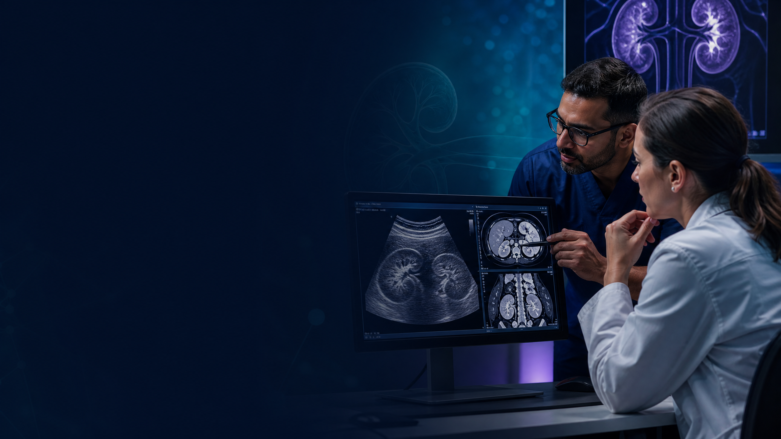
Visual learning: Chapter 38: Imaging in Nephrology – Complete Educational Package

Presentation resource: The Kidney Hub clinical-series PowerPoint for Chapters 31–40 accompanies these chapters for teaching use.