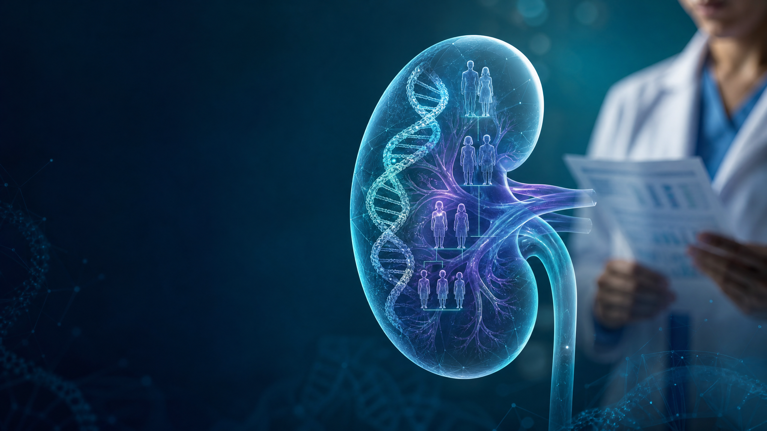
Chapter 23: Inherited Kidney Diseases
Learning Objectives
By the end of this chapter, learners will be able to:
– Explain the genetic basis and inheritance patterns of common inherited kidney diseases.
– Recognize key clinical features and extra-renal manifestations that suggest a hereditary kidney disorder.
– Apply a structured diagnostic approach, including indications and limitations of genetic testing.
– Outline disease-specific and general management strategies, including when to refer for specialist input.
– Provide core elements of genetic counseling and discuss reproductive options for affected families.
23.1 Introduction
Inherited kidney diseases account for a substantial proportion of chronic kidney disease (CKD) and end-stage kidney disease (ESKD) across the lifespan. Advances in next-generation sequencing (NGS) have transformed diagnosis, refined prognostication, and enabled targeted therapy in selected conditions. Many disorders are systemic, with extra-renal involvement (e.g., liver, ear, eye, cardiovascular, neurologic), and display variable penetrance and expressivity. Recognition of characteristic clinical patterns and judicious use of genetic testing improve diagnostic yield, guide family counseling, and may alter management.
Key inheritance patterns:
– Autosomal dominant (AD): vertical transmission; variable expressivity (e.g., ADPKD, ADTKD, some forms of FSGS).
– Autosomal recessive (AR): often pediatric onset (e.g., ARPKD, nephronophthisis, primary hyperoxaluria).
– X-linked: typically more severe in males (e.g., Alport syndrome, Fabry disease).
– Mitochondrial and oligogenic inheritance occur but are less common.
23.2 Common Inherited Kidney Disorders
1) Autosomal Dominant Polycystic Kidney Disease (ADPKD)
– Genetics: PKD1 (~85%) and PKD2 (~15%) variants; PKD1 tends to more rapid progression.
– Clinical: Progressive bilateral renal cysts, kidney enlargement, hypertension, hematuria, cyst infection, nephrolithiasis, pain.
– Extra-renal: Liver cysts, intracranial aneurysms (screening considered in high-risk), cardiac valve disease, abdominal wall hernias.
– Course: Heterogeneous; risk stratification informed by age-adjusted total kidney volume (Mayo Imaging Classification), genotype, and clinical trajectory.
2) Autosomal Recessive Polycystic Kidney Disease (ARPKD)
– Genetics: PKHD1 variants.
– Clinical: Enlarged echogenic kidneys in utero/infancy; congenital hepatic fibrosis and portal hypertension; pulmonary hypoplasia in severe neonatal disease.
3) Alport Syndrome
– Genetics: Type IV collagen genes (COL4A5 X-linked; COL4A3/4 AD or AR forms).
– Clinical: Persistent microscopic hematuria, proteinuria, progressive CKD; sensorineural hearing loss; ocular features (e.g., anterior lenticonus, dot-and-fleck retinopathy).
– Notes: Males with X-linked disease are typically more severely affected; carriers may have hematuria and variable CKD risk.
4) Fabry Disease
– Genetics: X-linked deficiency of alpha-galactosidase A (GLA gene) with accumulation of globotriaosylceramide (Gb3/GL-3).
– Clinical: Proteinuria and CKD (podocytopathy), neuropathic pain (acroparesthesias), angiokeratomas, hypohidrosis, corneal verticillata, cardiomyopathy, stroke.
– Diagnosis: Enzyme activity testing (reliable in males), lyso-Gb3; genetic testing essential in females.
5) Nephronophthisis (NPHP) and Autosomal Dominant Tubulointerstitial Kidney Disease (ADTKD)
– Nephronophthisis (AR): Tubulointerstitial nephropathy with corticomedullary cysts, salt wasting, polyuria/polydipsia; ESKD often in childhood/adolescence. Extra-renal features (retinal degeneration, cerebellar anomalies) in syndromic forms (ciliopathies).
– ADTKD (formerly “medullary cystic kidney disease”): AD tubulointerstitial disease due to UMOD, MUC1, REN, HNF1B, and others. Presents with gout/hyperuricemia (UMOD/REN), bland urinary sediment, progressive CKD; kidney size often normal or small.
6) Primary Hyperoxaluria (PH)
– Genetics: Enzyme defects in hepatic glyoxylate metabolism (e.g., AGXT in PH1) leading to overproduction of oxalate.
– Clinical: Nephrolithiasis, nephrocalcinosis, progressive CKD; systemic oxalosis in advanced disease.
– Therapeutics: Hydration, citrate, pyridoxine responsiveness in PH1 (subset), and RNA interference therapies for specific types; transplant strategies individualized.
7) Hereditary Renal Tubular Disorders
– Distal (type 1) and proximal (type 2) renal tubular acidosis: Hypokalemia, nephrocalcinosis/nephrolithiasis (especially distal RTA), growth issues in children.
– Gitelman/Bartter syndromes: Salt wasting, metabolic alkalosis, hypokalemia; nephrocalcinosis more typical in Bartter.
– Dent disease (X-linked): Low-molecular-weight proteinuria, hypercalciuria, nephrocalcinosis, CKD.
8) Congenital Anomalies of the Kidney and Urinary Tract (CAKUT)
– Spectrum includes renal agenesis, hypodysplasia, duplex systems, posterior urethral valves, and vesicoureteral reflux. Genetic contributors include HNF1B and others.
– Major cause of pediatric CKD; requires multidisciplinary follow-up.
9) Hereditary FSGS and Related Podocytopathies
– Genes include NPHS2, ACTN4, TRPC6, INF2, and others; typically steroid-resistant; recurrence risk after transplant is low in confirmed monogenic forms.
23.3 Patterns of Kidney Involvement
- Cystic disease: ADPKD, ARPKD, nephronophthisis, some ADTKD forms.
- Glomerular disease: Alport syndrome (GBM defect), Fabry disease (podocytopathy), hereditary FSGS.
- Tubulointerstitial disease: NPHP, ADTKD, primary hyperoxaluria (interstitial deposition), nephronophthisis-related ciliopathies.
- Nephrolithiasis/nephrocalcinosis: Primary hyperoxaluria, distal RTA, Dent disease, some Bartter variants.
- Electrolyte/acid–base disorders: RTA, Gitelman/Bartter.
23.4 Diagnostic Approach
1) History and Examination
– Detailed three-generation family history (CKD, dialysis/transplant, hearing/visual problems, gout, stones, aneurysms).
– Age at onset, growth/development, polyuria/polydipsia, pain, infections, extrarenal features (dermatologic, neurologic, ocular, hepatic).
2) Laboratory Testing
– Urinalysis (hematuria, proteinuria; low-molecular-weight proteinuria in Dent disease).
– Serum creatinine and eGFR using an age-appropriate equation; cystatin C may assist in select cases.
– Electrolytes, acid–base status; urinary electrolytes in tubulopathies.
– Disease-specific tests when indicated: enzyme activity and lyso-Gb3 for Fabry (especially in males); plasma/urine oxalate for PH; uric acid for ADTKD-UMOD/REN.
3) Imaging
– Renal ultrasound for kidney size, echotexture, cysts, nephrocalcinosis, and CAKUT features.
– MRI for ADPKD total kidney volume assessment when appropriate.
– Non-contrast MR angiography or CT angiography for intracranial aneurysm screening in ADPKD only in selected high-risk individuals (e.g., family history of aneurysm/SAH, prior aneurysm, high-risk occupations, or patient preference after counseling).
4) Kidney Biopsy
– Consider when noninvasive evaluation is non-diagnostic and histology may guide management (e.g., unexplained proteinuric disease).
– In Alport syndrome, electron microscopy may show GBM lamellation; immunostaining for alpha-5(IV) chain can support diagnosis.
– Note: Histology is often non-specific in genetic diseases and does not replace genetic testing.
5) Genetic Testing
– Pre-test counseling and informed consent are essential; discuss potential secondary findings and implications for relatives.
– Targeted single-gene testing for strong clinical suspicions (e.g., COL4A5 in classic X-linked Alport; PKD1/2 in typical ADPKD).
– Multigene panels for overlapping phenotypes (e.g., hereditary FSGS, tubulopathies).
– Exome/genome sequencing for undiagnosed or syndromic cases.
– Post-test counseling with cascade testing for at-risk relatives when a pathogenic/likely pathogenic variant is found.
– Limitations: Variants of uncertain significance are common; a negative test does not exclude a genetic etiology.
6) When to Refer
– Pediatric-onset or familial CKD; syndromic features; atypical presentations; consideration of disease-specific therapies; reproductive counseling; donor evaluation in related living donation.
23.5 Management and Genetic Counseling
A) General Renoprotective Measures
– Blood pressure management per local CKD and comorbidity guidelines; RAAS blockade preferred when proteinuria is present and tolerated.
– Lifestyle measures: sodium restriction, weight management, smoking cessation, exercise.
– Avoid nephrotoxins where possible (e.g., NSAIDs, repeated iodinated contrast without indication).
B) Disease-Specific Strategies
– ADPKD:
– Candidate selection for tolvaptan should be based on evidence of rapid progression (e.g., Mayo class 1C–1E, early-onset hypertension or urologic complications, genotype/PROPKD score, or historical eGFR decline). Follow local eligibility criteria and monitor liver function regularly. Counsel on aquaretic side effects.
– Manage complications: pain (stepwise approach; consider cyst decompression for selected large cysts), hematuria, cyst infection (lipid-soluble antibiotics), nephrolithiasis (hydration, stone-directed therapy).
– Intracranial aneurysm screening for high-risk groups as above; interval and modality per local practice.
– ARPKD:
– Multidisciplinary care with attention to blood pressure, growth/nutrition, portal hypertension and biliary complications, and respiratory issues in infancy.
– Alport Syndrome:
– Early initiation of RAAS blockade to reduce proteinuria and slow CKD progression where appropriate.
– Audiology and ophthalmology assessments; hearing support including hearing aids/cochlear implant when indicated.
– Transplantation: excellent outcomes; rare post-transplant anti-GBM–like nephritis may occur, requiring close monitoring.
– Fabry Disease:
– Disease-specific therapy: enzyme replacement therapy in appropriate candidates; oral chaperone therapy for amenable GLA variants.
– Adjunctive RAAS blockade for proteinuria; manage cardiac and cerebrovascular risks; monitor lyso-Gb3 where available.
– Nephronophthisis:
– Supportive CKD care; manage polyuria with access to fluids; screen for and manage extra-renal features in syndromic forms; plan for kidney replacement therapy.
– ADTKD:
– General CKD care; treat gout/hyperuricemia when indicated; no disease-specific antifibrotic therapy available.
– Primary Hyperoxaluria:
– High fluid intake, citrate therapy, dietary measures; pyridoxine trial in PH1; RNA interference therapies available for specific types (per local availability).
– Advanced disease may require combined or staged organ transplantation strategies individualized to type and response.
– Hereditary RTA and Tubulopathies:
– Alkali therapy for RTA; potassium repletion as needed; thiazides for hypercalciuria in distal RTA.
– Salt and magnesium repletion for Gitelman; tailored therapy for Bartter variants.
– Dent disease: stone prevention strategies, thiazides for hypercalciuria (if tolerated), CKD care.
C) Kidney Replacement Therapy and Transplant Considerations
– Transplantation is preferred for ESKD in most inherited diseases.
– Recurrence risk:
– Low in monogenic FSGS compared with idiopathic forms.
– Primary hyperoxaluria requires oxalate management; systemic oxalosis can affect graft outcomes.
– ADPKD does not recur in the graft.
– Living Donor Evaluation:
– Related donors require careful assessment for shared genetic risk; consider genetic testing when a familial pathogenic variant is known or strongly suspected.
– APOL1 risk variants in potential donors of sub-Saharan African ancestry may inform counseling per local practice.
D) Genetic Counseling and Reproductive Options
– Provide counseling on inheritance pattern, penetrance/expressivity, and recurrence risks.
– Offer cascade testing for at-risk relatives when a familial pathogenic variant is identified.
– Discuss reproductive options, including prenatal diagnosis and preimplantation genetic testing for monogenic disorders, in accordance with local regulations and patient values.
SUMMARY
- Inherited kidney diseases are a major cause of CKD/ESKD in both pediatric and adult populations, often with multisystem involvement.
- Recognizable clinical patterns (e.g., cystic kidneys, hematuria with hearing loss, nephrolithiasis/nephrocalcinosis, tubulointerstitial disease with gout) guide targeted evaluation.
- Genetic testing, when coupled with thoughtful phenotyping and counseling, improves diagnostic certainty and informs management; however, results may be inconclusive, and clinical judgment remains essential.
- Disease-specific therapies exist for selected conditions (e.g., tolvaptan for rapidly progressive ADPKD; enzyme/chaperone therapy in Fabry; targeted therapies in primary hyperoxaluria).
- Early renoprotective measures, multidisciplinary care, and timely transplant planning optimize outcomes.
- Genetic counseling, cascade testing, and informed reproductive discussions are integral to patient and family care.
CLINICAL PEARLS
- Obtain a three-generation family history in all patients with early-onset CKD, persistent hematuria/proteinuria, bilateral cysts, or syndromic features.
- Persistent microscopic hematuria with hearing loss suggests Alport syndrome; refer for genetic testing of COL4A3/4/5.
- In males, low alpha-galactosidase A activity strongly supports Fabry disease; females require genetic testing due to lyonization.
- In ADPKD, risk stratify progression using age-adjusted total kidney volume and clinical/genetic factors to guide tolvaptan eligibility.
- Distal RTA often presents with nephrocalcinosis and hypokalemia; alkali therapy reduces stone risk and preserves growth in children.
- Consider ADTKD in families with bland urine sediment, gout/hyperuricemia, and AD CKD; cysts may be minimal or absent.
- Genetic FSGS has a low risk of post-transplant recurrence; confirm monogenic etiology when possible before listing.
- Screen selectively for intracranial aneurysm in ADPKD based on risk factors and shared decision-making.
- Related living donor candidates from affected families often need genetic evaluation before approval.
VISUAL MATERIALS
Proposed figures and tables (to be developed):
– Diagram: Inheritance patterns (AD, AR, X-linked) with example kidney disorders.
– Flowchart: Diagnostic pathway for suspected inherited kidney disease, including when to order targeted gene tests vs panels vs exome/genome.
– Schematic: ADPKD pathophysiology and progression; depiction of Mayo Imaging Classification concept.
– Table: Distinguishing features of ADPKD vs ARPKD vs NPHP vs ADTKD (age at onset, imaging, urine findings, extrarenal signs).
– Infographic: Alport syndrome—GBM changes, hearing/ocular features, testing strategy.
– Table: Disease-specific therapies (e.g., tolvaptan, Fabry ERT/chaperone, PH therapies) with indications and key monitoring points.
– Diagram: Stone-forming genetic disorders (PH, distal RTA, Dent) with prevention strategies.
MULTIPLE CHOICE QUESTIONS
1) Which is the most common inherited kidney disease?
– A) Alport syndrome
– B) Autosomal dominant polycystic kidney disease (ADPKD)
– C) Nephronophthisis
– D) Fabry disease
Answer: B) Autosomal dominant polycystic kidney disease (ADPKD)
Explanation: ADPKD is the most prevalent hereditary kidney disorder worldwide.
2) A 15-year-old boy has persistent microscopic hematuria, progressive hearing loss, and a maternal uncle with ESKD. Most likely diagnosis?
– A) Alport syndrome
– B) ARPKD
– C) Fabry disease
– D) ADTKD
Answer: A) Alport syndrome
Explanation: Hematuria with X-linked family pattern and hearing loss is characteristic of Alport syndrome.
3) Which extra-renal manifestation is most closely associated with ADPKD?
– A) Cerebellar ataxia
– B) Intracranial aneurysm
– C) Angiokeratomas
– D) Anterior lenticonus
Answer: B) Intracranial aneurysm
Explanation: Intracranial aneurysms occur more frequently in ADPKD; screening is targeted to high-risk groups.
4) In males suspected of having Fabry disease, the most informative initial test is:
– A) Kidney biopsy
– B) Serum alpha-galactosidase A activity
– C) Serum complement levels
– D) Urinary oxalate
Answer: B) Serum alpha-galactosidase A activity
Explanation: Enzyme activity is typically low in affected males; females require genetic testing due to variable enzyme levels.
5) A young adult with CKD, early-onset gout, bland urine sediment, and an affected parent most likely has:
– A) Nephronophthisis
– B) ADTKD due to UMOD
– C) Primary hyperoxaluria
– D) Distal RTA
Answer: B) ADTKD due to UMOD
Explanation: AD tubulointerstitial disease with hyperuricemia/gout is classic for UMOD-related ADTKD.
6) Which statement about genetic testing in inherited kidney diseases is most accurate?
– A) A negative multigene panel excludes a genetic cause
– B) Exome sequencing always identifies a causal variant
– C) Variants of uncertain significance may require periodic reinterpretation
– D) Kidney biopsy is superior to genetic testing for diagnosis
Answer: C) Variants of uncertain significance may require periodic reinterpretation
Explanation: VUS findings are common; periodic review and segregation analysis may clarify significance.
7) Which patient profile most supports consideration of tolvaptan for ADPKD?
– A) Age 70 with stable eGFR and Mayo class 1A kidneys
– B) Age 25, PKD1-truncating variant, rapid eGFR decline, Mayo class 1D
– C) Age 60, PKD2 variant, no hypertension, Mayo class 1B
– D) Age 40 with nephronophthisis
Answer: B) Age 25, PKD1-truncating variant, rapid eGFR decline, Mayo class 1D
Explanation: Evidence of rapid progression and high-risk features favor tolvaptan consideration per local criteria.
8) A child with nephrocalcinosis, hypokalemia, and normal anion gap metabolic acidosis most likely has:
– A) Distal renal tubular acidosis
– B) Gitelman syndrome
– C) Bartter syndrome type I
– D) Nephronophthisis
Answer: A) Distal renal tubular acidosis
Explanation: dRTA causes non–anion gap metabolic acidosis, hypokalemia, and predisposes to nephrocalcinosis.
9) In primary hyperoxaluria type 1, which therapy may reduce oxalate production in a subset of patients?
– A) Prednisone
– B) Pyridoxine (vitamin B6)
– C) Furosemide
– D) Allopurinol
Answer: B) Pyridoxine (vitamin B6)
Explanation: Some PH1 patients with specific AGXT variants respond to pyridoxine with reduced oxalate production.
10) Which statement about post-transplant recurrence risk is correct?
– A) ADPKD invariably recurs in the allograft
– B) Monogenic FSGS generally has a low recurrence risk
– C) Alport syndrome often recurs and rapidly destroys the graft
– D) Primary hyperoxaluria does not affect the kidney allograft
Answer: B) Monogenic FSGS generally has a low recurrence risk
Explanation: Genetic FSGS has a low risk of recurrence; ADPKD does not recur, while PH can injure the graft due to systemic oxalate.
POWERPOINT PRESENTATION
Slide 1: Title — Inherited Kidney Diseases: Recognition, Diagnosis, and Management
– Speaker points: Scope; impact on CKD/ESKD; multisystem nature; goals of session.
Slide 2: Inheritance Patterns and Epidemiology
– Speaker points: AD/AR/X-linked; penetrance/expressivity; pediatric vs adult onset.
Slide 3: Cystic Diseases — ADPKD and ARPKD
– Speaker points: Genetics (PKD1/2; PKHD1); imaging; extra-renal features; progression risk tools.
Slide 4: Glomerular Disorders — Alport and Fabry
– Speaker points: COL4A3/4/5 phenotypes; hearing/ocular signs; Fabry systemic clues; testing strategies.
Slide 5: Tubulointerstitial Diseases — NPHP and ADTKD
– Speaker points: NPHP ciliopathy features; ADTKD genes (UMOD, MUC1, REN, HNF1B); clinical hallmarks.
Slide 6: Stone-Formers and Tubulopathies
– Speaker points: Primary hyperoxaluria; distal RTA; Dent disease; key labs and prevention.
Slide 7: Diagnostic Algorithm
– Speaker points: Family history; targeted labs/imaging; when to biopsy; genetic testing tiers; counseling.
Slide 8: Disease-Specific Therapies
– Speaker points: Tolvaptan selection/monitoring; Fabry ERT/chaperone; PH therapies; supportive CKD care.
Slide 9: Transplant and Donor Considerations
– Speaker points: Recurrence risks; Alport post-transplant issues; related donor genetic evaluation; APOL1 counseling.
Slide 10: Genetic Counseling and Reproductive Options
– Speaker points: Inheritance/risk; cascade testing; prenatal and preimplantation testing; ethical considerations.
Slide 11: Clinical Pearls and Pitfalls
– Speaker points: Red flags; avoiding overreliance on biopsy; interpreting VUS; multidisciplinary care.
Slide 12: Summary and Take-Home Messages
– Speaker points: Early recognition; targeted testing; individualized therapy; family-centered care.
Educational disclaimer: This chapter is for educational purposes for healthcare professionals. Management decisions should be individualized and aligned with local guidelines, patient comorbidities, and specialist consultation.
Visual learning: Inherited Kidney Diseases

Presentation resource: The Kidney Hub clinical-series PowerPoint for Chapters 22–30 accompanies these chapters for teaching use.