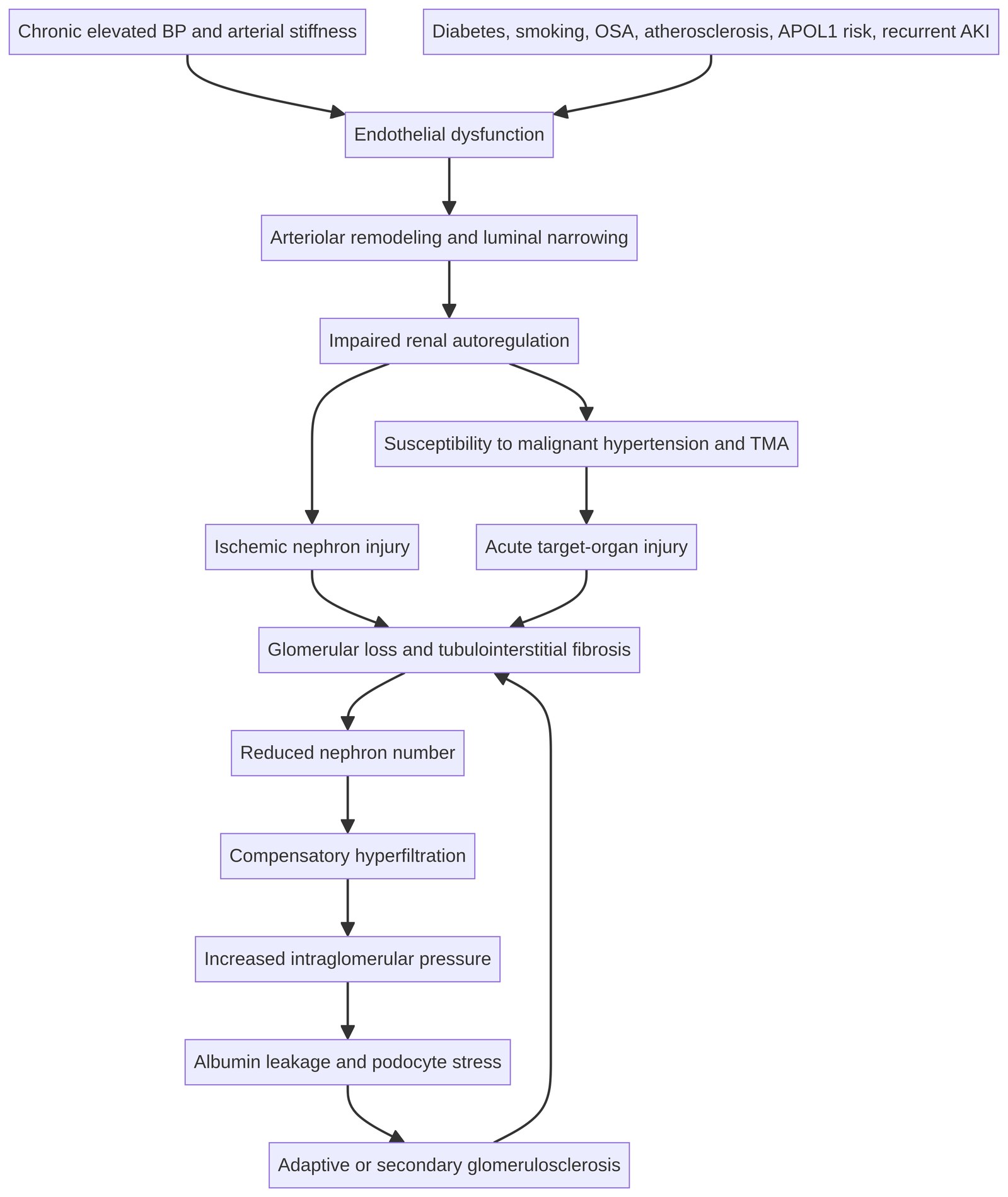
Specialist Chapter for Nephrologists
Clinical scope. Hypertension and CKD are bidirectionally related. “Hypertensive nephrosclerosis” should be treated as a probability-based clinicopathologic diagnosis, not as an automatic explanation for every patient with hypertension and reduced eGFR. Atypical urine findings, disproportionate proteinuria, rapid progression, systemic disease, or acute kidney injury should prompt a broader diagnostic evaluation.
Chapter Summary
Hypertension-associated kidney disease encompasses a spectrum ranging from chronic arterionephrosclerosis to malignant hypertension with acute endothelial injury and thrombotic microangiopathy. The clinical phenotype is usually characterized by long-standing hypertension, slowly declining kidney function, relatively bland urine sediment, and low-to-moderate albuminuria; however, these features are neither sufficiently sensitive nor specific to establish causality. Diabetes, aging-related nephron loss, APOL1-associated disease, primary glomerular disease, renovascular disease, chronic tubulointerstitial disease, and medication-related injury may coexist or be misclassified as hypertensive kidney disease.
Management should combine accurate blood-pressure phenotyping, sodium and volume assessment, risk-based RAAS blockade, SGLT2 inhibition when indicated, treatment of resistant hypertension, cardiovascular-risk reduction, and timely kidney-failure planning. The KDIGO systolic blood-pressure target of less than 120 mmHg applies to standardized office measurement and only when tolerated; it should not be transposed directly to casual clinic readings.[1] [2]
Learning Objectives
After completing this chapter, the learner should be able to:
- Distinguish hypertension-associated kidney disease, arterionephrosclerosis, malignant hypertension, and hypertensive emergency.
- Describe the vascular, glomerular, tubular, inflammatory, and genetic mechanisms of hypertension-related kidney injury.
- Obtain and interpret standardized office, home, and ambulatory blood-pressure data.
- Recognize clinical features that are atypical for presumed hypertensive nephrosclerosis.
- Decide when serologic testing, renovascular evaluation, genetic assessment, or kidney biopsy is appropriate.
- Construct a treatment plan integrating sodium reduction, volume control, ACE inhibitor or ARB therapy, SGLT2 inhibition, and resistant-hypertension management.
- Identify malignant hypertension and kidney-predominant thrombotic microangiopathy requiring urgent monitored care.
- Use eGFR trajectory, albuminuria, KFRE, comorbidity, and patient goals to guide kidney-failure preparation.
Related Kidney Hub resources. Continue with the Kidney Hub chapter library, explore interactive nephrology cases, and review the guideline library for related clinical topics.
12.1 Terminology and Diagnostic Framing
Hypertension-associated kidney disease is a clinical category in which chronic blood-pressure exposure is judged to contribute materially to kidney damage. Arterionephrosclerosis is a morphologic description that may include arteriolar hyalinosis, arterial intimal thickening, ischemic glomerular injury, global glomerulosclerosis, tubular atrophy, and interstitial fibrosis. These lesions are common in aging, diabetes, atherosclerosis, and other kidney diseases; they are therefore not specific for hypertension.
A patient may develop hypertension as a consequence of CKD, develop CKD as a consequence of hypertension, or have both conditions driven by another process. Causal inference should integrate the chronology of blood pressure and eGFR, albuminuria, urine sediment, kidney size, diabetes status, vascular disease, medication exposure, and the presence or absence of systemic features.
The traditional clinical phenotype of presumed hypertensive nephrosclerosis includes many years of hypertension, slowly progressive eGFR loss, bland sediment, low-grade albuminuria, and small or normal-sized kidneys. This phenotype increases the probability of arterionephrosclerosis but does not exclude other disease. Clinical criteria alone may have limited sensitivity for biopsy-defined lesions, particularly in patients with APOL1 risk variants or coexisting glomerular disease.[5] [6]
12.2 Pathophysiology
Chronic pressure load causes endothelial dysfunction, vascular smooth-muscle remodeling, increased arterial stiffness, impaired autoregulation, and progressive narrowing of small renal vessels. Reduced perfusion produces ischemic nephron loss, while surviving nephrons develop compensatory hyperfiltration and increased intraglomerular pressure. This combination promotes podocyte stress, albumin leakage, glomerulosclerosis, tubular atrophy, and interstitial fibrosis.
Vascular Injury
Hyaline arteriolosclerosis reflects plasma-protein leakage and vascular smooth-muscle injury. Fibroelastic intimal thickening and medial hypertrophy reduce luminal caliber and impair pressure transmission. The severity of vascular disease may be amplified by diabetes, smoking, dyslipidemia, systemic atherosclerosis, sleep apnea, and recurrent episodes of acute hypertension.
Glomerular and Tubulointerstitial Injury
Ischemic glomeruli may become globally sclerosed, while remaining glomeruli may enlarge and hyperfilter. Focal segmental sclerosis can develop as an adaptive lesion and may be associated with substantial proteinuria. Tubular atrophy and interstitial fibrosis are major histologic correlates of irreversible kidney-function loss, regardless of the initiating vascular insult.
Malignant Hypertension
Abrupt severe blood-pressure elevation can produce endothelial injury, fibrinoid necrosis, hyperplastic arteriolosclerosis, and a kidney-predominant thrombotic microangiopathy. The clinical syndrome may include rapidly rising creatinine, hematuria, proteinuria, thrombocytopenia, microangiopathic hemolysis, retinal hemorrhages or papilledema, encephalopathy, pulmonary edema, or heart failure. Malignant hypertension must be evaluated as an acute target-organ syndrome rather than described merely as “uncontrolled hypertension.”
APOL1-Associated Risk
APOL1 G1 and G2 risk variants are associated with FSGS, HIV-associated nephropathy, collapsing lesions, and hypertension-attributed kidney failure. Risk is modified by environmental and biologic “second hits,” and most individuals with high-risk genotypes do not develop kidney disease. Genetic ancestry should not be used as a proxy for genotype, and race-based assumptions should not be used to assign a kidney diagnosis. Testing may be considered when the result can affect counseling, transplant assessment, research participation, or interpretation of an atypical presentation.[5]
12.3 Clinical Phenotypes and Differential Diagnosis
| Clinical phenotype | Features that support hypertension-associated disease | Diagnoses that must remain in the differential |
|---|---|---|
| Chronic, slowly progressive CKD | Long-standing hypertension, bland sediment, low-to-moderate albuminuria, gradual eGFR decline | Diabetes, aging-related CKD, chronic tubulointerstitial disease, APOL1-associated disease |
| Albuminuric CKD | Persistent albuminuria with hypertension and progressive eGFR loss | Diabetic kidney disease, FSGS, IgA nephropathy, membranous nephropathy, obesity-related glomerulopathy |
| Rapid decline or AKI | Recent severe hypertension, abrupt creatinine increase, volume or medication trigger | TMA, vasculitis, acute GN, interstitial nephritis, obstruction, renal artery disease |
| Malignant-hypertension syndrome | Severe BP elevation with retinal, neurologic, cardiac, or renal target-organ injury | Scleroderma renal crisis, pheochromocytoma, TTP, complement-mediated TMA, acute GN |
| Resistant hypertension | Uncontrolled BP on three agents including a diuretic, or control requiring four or more agents | Primary aldosteronism, renal artery disease, OSA, drug-induced hypertension, volume excess |
| Non-albuminuric eGFR decline | Low ACR with vascular disease, asymmetric kidneys, or ischemic features | Ischemic nephropathy, obstruction, tubulointerstitial disease, medication toxicity |
12.4 Blood-Pressure Phenotyping
Blood-pressure management begins with reliable measurement. For standardized office measurement, the patient should rest quietly, use an appropriate cuff, sit with back supported and feet on the floor, keep the arm supported at heart level, avoid conversation, and undergo repeated measurements. The clinician should document the technique because a target derived from standardized measurement cannot be safely mapped to an arbitrary casual reading.
Home blood-pressure monitoring is useful for longitudinal titration and for identifying white-coat or masked hypertension. Ambulatory monitoring adds information about nocturnal hypertension, dipping status, morning surge, and the relationship between symptoms and blood pressure. Discordance between office and out-of-office readings should be resolved before intensifying treatment, particularly in older adults, patients with orthostasis, and patients at risk of falls.
The KDIGO target systolic blood pressure of less than 120 mmHg is suggested for adults with CKD and high blood pressure when tolerated and when measured using standardized office technique.[1] Individualization is required for symptomatic orthostasis, frailty, limited life expectancy, advanced autonomic dysfunction, or inability to reproduce standardized measurement.[2]
12.5 Diagnostic Evaluation
The diagnostic work-up should establish chronicity, quantify kidney damage, identify complications, assess secondary hypertension, and test whether the presumed diagnosis is sufficiently coherent to avoid biopsy.
| Domain | Initial assessment | Findings that should change the pathway |
|---|---|---|
| Blood pressure | Standardized office readings, home log, or ABPM | Marked variability, nocturnal hypertension, orthostasis, or resistant hypertension |
| Kidney function | Creatinine/eGFR trend, electrolytes, bicarbonate, calcium, phosphate as indicated | Rapid eGFR decline, AKI, hyperkalemia, acidosis, or unexplained electrolyte disorder |
| Urine | Urinalysis with microscopy, UACR, and protein quantification when needed | Dysmorphic RBCs, RBC casts, granular casts, nephrotic-range proteinuria, or active sediment |
| Structure | Ultrasound for kidney size, echogenicity, asymmetry, cysts, and obstruction | Marked asymmetry, obstruction, unexpected enlargement, or complex cystic disease |
| Systemic injury | Fundoscopy, CBC, platelet count, hemolysis profile, ECG, cardiac assessment, neurologic examination | Retinopathy, TMA, pulmonary edema, heart failure, encephalopathy, or focal neurologic signs |
| Secondary causes | Medication and supplement review; targeted endocrine, sleep-apnea, and renovascular evaluation | Hypokalemia, abrupt onset, episodic symptoms, abdominal bruit, or flash pulmonary edema |
The level of albuminuria is especially important. Low albuminuria supports—but does not prove—a vascular or tubulointerstitial process. Heavy or rapidly increasing proteinuria should prompt consideration of glomerular disease, adaptive FSGS, APOL1-associated disease, or a combined process.
12.6 When to Pursue Further Testing or Kidney Biopsy
Kidney biopsy should be considered when the result is likely to change treatment, prognosis, genetic counseling, or transplant planning. Indications include substantial or rapidly increasing proteinuria, active urine sediment, rapid or unexplained eGFR decline, unexplained AKI, systemic disease, unexpected kidney size, suspected TMA, or a phenotype that is not adequately explained by hypertension.
A biopsy showing arterionephrosclerosis does not establish that hypertension was the sole cause of CKD. Histology should be interpreted with the clinical chronology, degree and type of proteinuria, urine microscopy, diabetes status, vascular disease, possible APOL1 risk, and the presence of primary or secondary glomerular lesions.
In suspected malignant hypertension with thrombocytopenia or hemolysis, obtain a blood smear, LDH, haptoglobin, bilirubin, reticulocyte count, coagulation studies, and urinalysis. ADAMTS13, complement evaluation, autoimmune testing, and kidney biopsy should be selected according to the clinical context. Life-threatening target-organ injury requires urgent treatment and should not await biopsy results.
Sodium, Weight, Activity, and Volume
A dietary sodium intake below 2 g per day is recommended for many adults with CKD when feasible, but implementation should account for food access, cultural patterns, comorbid heart failure, nutritional status, and patient preferences.[1] Review processed foods, restaurant meals, sodium-containing medications, and dietary practices that produce large day-to-day changes in sodium balance.
Assess extracellular volume using the examination, weight trajectory, edema, pulmonary findings, orthostatic symptoms, diuretic exposure, heart failure status, and residual kidney function. Diuretic choice and dose should reflect eGFR, sodium intake, edema, and response. Fluid restriction should not be prescribed automatically without a specific indication such as clinically important hyponatremia or severe congestion.
RAAS Blockade
In CKD with hypertension and albuminuria, an ACE inhibitor or ARB should generally be used and titrated to the highest tolerated approved dose.[1] Monitor creatinine and potassium after initiation and dose escalation. An early hemodynamic change in eGFR may occur, but a large or progressive creatinine rise requires evaluation for volume depletion, NSAIDs, obstruction, excessive diuresis, renal artery disease, or another acute process.
Do not combine an ACE inhibitor with an ARB or a direct renin inhibitor for routine CKD treatment. Hyperkalemia should trigger medication review, correction of reversible factors, and consideration of potassium-lowering strategies when appropriate rather than automatic discontinuation of a strongly indicated therapy.
SGLT2 Inhibition
SGLT2 inhibitors reduce kidney and heart-failure risk in many eligible patients with CKD, with and without diabetes, according to the specific agent’s prescribing criteria and current guideline recommendations.[2] A small early eGFR dip may reflect a hemodynamic effect. Before and during treatment, assess volume status, genital-infection risk, ketoacidosis risk, kidney function, and the need to hold therapy during prolonged fasting, severe acute illness, or major surgery.
Resistant Hypertension
Confirm that resistant hypertension is genuine. Review standardized and out-of-office measurements, adherence, medication access, dosing schedule, sodium intake, volume status, sleep apnea, NSAIDs, glucocorticoids, stimulants, oral contraceptives, calcineurin inhibitors, erythropoiesis-stimulating agents, and licorice exposure.
A common multidrug framework includes a RAAS blocker when indicated, a long-acting dihydropyridine calcium-channel blocker, and an effective diuretic. Additional therapy should be guided by volume status, potassium, heart rate, coronary disease, heart failure, and suspected secondary causes. Mineralocorticoid receptor antagonism may be useful in selected patients but requires careful potassium and kidney-function monitoring.
| Treatment problem | Practical nephrology response |
|---|---|
| Apparent resistant hypertension | Confirm technique, adherence, access, white-coat effect, and medication reconciliation. |
| Volume-mediated hypertension | Reassess sodium intake, edema, heart failure, diuretic dose, and kidney function. |
| Hyperkalemia limiting RAAS blockade | Remove reversible contributors, adjust diet appropriately, consider potassium binders, and reassess the risk-benefit balance. |
| Orthostatic symptoms | Measure standing BP, review diuretics and vasodilators, and individualize the BP target. |
| Rapidly worsening BP | Evaluate for malignant hypertension, TMA, renal artery disease, endocrine causes, medication exposure, and AKI. |
12.8 Malignant Hypertension and Hypertensive Emergency
A hypertensive emergency is severe blood-pressure elevation accompanied by acute target-organ injury. Kidney manifestations include rapidly rising creatinine, hematuria, proteinuria, oliguria, TMA, and pulmonary edema. Evaluate neurologic, cardiac, retinal, aortic, and renal injury in parallel.
Treatment generally requires monitored care and a titratable intravenous agent when acute organ injury is present. The rate and magnitude of reduction depend on the organ syndrome; precipitous normalization may compromise cerebral, coronary, or renal perfusion. Coordinate nephrology, critical care, cardiology, neurology, ophthalmology, and hematology input when indicated.
12.9 Secondary Hypertension and Renovascular Disease
Consider secondary hypertension with abrupt onset, severe or early-onset disease, resistant hypertension, hypokalemia, episodic headache or palpitations, disproportionate kidney dysfunction, asymmetric kidneys, recurrent flash pulmonary edema, or a marked creatinine change after RAAS blockade.
Renal artery stenosis may result from atherosclerosis or fibromuscular dysplasia. Imaging should be selected according to kidney function, contrast risk, anatomy, local expertise, and whether the result is likely to change treatment. Most stable atherosclerotic disease is managed medically. Revascularization is reserved for selected high-risk presentations, such as recurrent flash pulmonary edema or difficult-to-control hypertension with compelling anatomic and clinical features, after multidisciplinary review.
12.10 Cardiovascular Risk and Comorbidity
CKD with hypertension is a high cardiovascular-risk state. Management should address lipid disorders, smoking, diabetes, obesity, sleep apnea, physical activity, vaccination, atrial fibrillation, heart failure, and avoidance of nephrotoxins. The treatment plan should not focus on a blood-pressure number while overlooking albuminuria, volume status, glycemic risk, or established cardiovascular disease.
12.11 Prognosis and Kidney-Failure Planning
Prognosis should be based on eGFR, UACR, eGFR slope, episodes of AKI, KFRE when applicable, cardiovascular disease, frailty, age, comorbidity, and patient goals. Hypertension-associated CKD may progress slowly, but heavy proteinuria, malignant hypertension, recurrent AKI, APOL1-associated disease, and persistent uncontrolled blood pressure may accelerate decline.
Transplant and dialysis education should begin early enough to allow informed choice. Discuss living and deceased-donor transplantation, peritoneal dialysis, home hemodialysis, in-center hemodialysis, conservative kidney management, vascular access planning, and advance-care planning. The timing should be individualized rather than determined by eGFR alone.
12.12 Clinical Pearls
- Hypertension plus CKD is not synonymous with hypertensive nephrosclerosis.
- Standardized blood pressure measurement is a prerequisite for interpreting an intensive systolic target.
- Heavy proteinuria, active sediment, rapid decline, or systemic features should trigger a broader differential.
- Arterionephrosclerosis on biopsy may be contributory, coincident, or secondary rather than the sole cause of CKD.
- Treat sodium excess and volume expansion before adding layers of antihypertensive medication.
- Recheck creatinine and potassium after RAAS-blocker initiation or dose escalation.
- A modest early eGFR dip after SGLT2 inhibition can be hemodynamic; severe or progressive decline requires assessment.
- Malignant hypertension with hemolysis or thrombocytopenia is a medical emergency and may represent TMA.
- Use genetic testing selectively and do not substitute ancestry or race for APOL1 genotype.
- Kidney-failure planning should begin before urgent dialysis decisions are required.
12.13 Diagnostic and Treatment Pathway
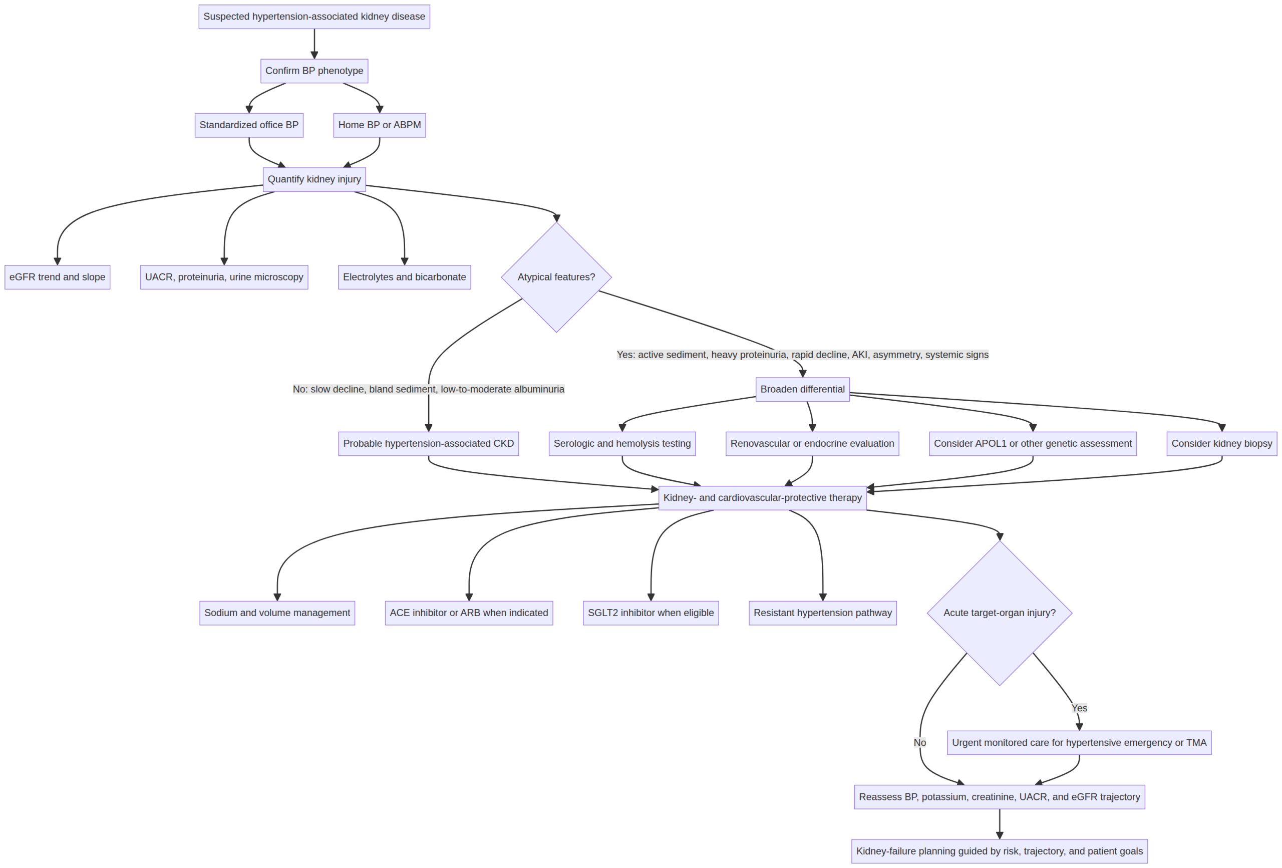
Figure 12.2. Diagnostic and treatment pathway for suspected hypertension-associated kidney disease.
Begin by confirming the blood-pressure phenotype with standardized office measurement and, when needed, home monitoring or ABPM. Quantify kidney injury using eGFR trajectory, UACR, urinalysis with microscopy, and structural assessment. If the presentation is atypical—heavy or rapidly increasing proteinuria, active sediment, rapid eGFR loss, systemic features, asymmetric kidneys, or suspected secondary hypertension—broaden the differential and consider targeted testing or kidney biopsy when the result could change management.
Patients with a coherent chronic vascular phenotype should receive kidney- and cardiovascular-protective therapy, including sodium and volume management, RAAS blockade when indicated, and SGLT2 inhibition when eligible. Any branch showing acute target-organ injury—encephalopathy, pulmonary edema, rapidly progressive AKI, retinal injury, myocardial injury, thrombocytopenia, or hemolysis—should proceed to monitored urgent care while TMA and other secondary causes are evaluated in parallel.
12.14 Clinical Safety Note
This chapter is an educational resource and does not replace individualized nephrology, hypertension, cardiology, critical-care, hematology, or transplant assessment. Antihypertensive and kidney-protective therapies may cause hypotension, acute kidney injury, hyperkalemia, electrolyte disorders, genital infections, ketoacidosis, and clinically important drug interactions. Suspected hypertensive emergency, malignant hypertension, TMA, pulmonary edema, encephalopathy, or rapidly progressive kidney injury requires urgent clinical evaluation.
Question 1 — Typical phenotype or premature closure?
Difficulty: Resident to nephrologist.
A 58-year-old patient has a 15-year history of hypertension, eGFR 42 mL/min/1.73 m², UACR 80 mg/g, bland urine sediment, normal potassium, and a gradual eGFR decline of approximately 2 mL/min/1.73 m² per year. Kidney ultrasound shows mildly small, symmetric kidneys without obstruction. What makes arterionephrosclerosis plausible, and what prevents diagnostic certainty?
Best answer
The phenotype is compatible with hypertension-associated CKD because the patient has long-standing hypertension, a slowly progressive course, low-to-moderate albuminuria, bland sediment, and symmetric small kidneys. These findings increase the probability of chronic vascular and ischemic injury. They do not prove that hypertension is the sole cause. Diabetes, atherosclerotic or ischemic disease, aging-related nephron loss, chronic tubulointerstitial disease, APOL1-associated disease, medication exposure, and an unrecognized glomerular process should still be considered.
Clinical reasoning
The first task is to establish whether the kidney disease is truly chronic and whether the eGFR slope is reliable. Review older creatinine values, intercurrent AKI episodes, NSAID or proton-pump-inhibitor exposure, volume depletion, urinary obstruction, and changes in muscle mass. A gradual slope is reassuring but should not be interpreted without confirming the quality and frequency of the measurements.
The urine phenotype is informative. An ACR of 80 mg/g represents moderately increased albuminuria, which is compatible with vascular disease but also occurs in diabetes, obesity-related glomerulopathy, adaptive FSGS, and early primary glomerular disease. Bland sediment lowers the probability of proliferative GN or active vasculitis but does not exclude all glomerular disease. Kidney size supports chronicity and vascular or ischemic injury, but small kidneys can also occur in reflux nephropathy, chronic interstitial disease, and advanced disease of many causes.
Appropriate evaluation and management
Confirm standardized and out-of-office blood pressure, quantify the eGFR trajectory and UACR periodically, review medications and sodium intake, and assess cardiovascular and retinal disease. If hypertension and albuminuria persist, an ACE inhibitor or ARB is generally appropriate when tolerated; an SGLT2 inhibitor may be appropriate according to current CKD eligibility criteria.[1] [2] The patient should be monitored for potassium, creatinine, orthostasis, and volume status.
Kidney biopsy is not automatically indicated when the phenotype is typical and the result is unlikely to change treatment. It becomes more reasonable if proteinuria increases substantially, the eGFR slope accelerates, hematuria develops, systemic features appear, or the clinical course becomes discordant with presumed arterionephrosclerosis.
Common pitfall
The common error is to treat the coexistence of hypertension and CKD as proof of causality. The correct formulation is: “The phenotype is most consistent with hypertension-associated CKD, while alternative and coexisting causes have been assessed and remain less likely.”
Question 2 — Atypical proteinuria and active sediment
Difficulty: Nephrologist.
A 62-year-old patient previously labeled as having hypertensive nephrosclerosis develops nephrotic-range proteinuria, dysmorphic hematuria, RBC casts, and a fall in eGFR from 48 to 28 mL/min/1.73 m² over four months. Blood pressure is 178/102 mmHg. What is the next step, and why is simply intensifying antihypertensive therapy inadequate?
Best answer
The diagnosis must be reopened urgently. The combination of nephrotic-range proteinuria, dysmorphic hematuria, RBC casts, and rapid eGFR decline is atypical for isolated arterionephrosclerosis and strongly suggests an active glomerular or systemic process. The patient requires urgent nephrology evaluation, targeted serologic testing, structural assessment, medication review, and consideration of kidney biopsy if the result will guide immunosuppression or other disease-specific treatment.
Clinical reasoning
Nephrotic-range proteinuria indicates substantial glomerular barrier injury or, less commonly, severe adaptive hyperfiltration injury. Dysmorphic RBCs and RBC casts localize the process to the glomerulus and make isolated chronic vascular nephropathy insufficient as an explanation. The rapid eGFR decline may represent crescentic GN, infection-related GN, membranoproliferative disease, lupus nephritis, ANCA-associated vasculitis, anti-GBM disease, severe IgA nephropathy, or a secondary FSGS pattern.
The initial evaluation should include repeat urinalysis with microscopy, quantitative protein measurement, CBC, electrolytes, albumin, lipid profile, complements, ANA, ANCA, anti-GBM antibody, hepatitis B and C testing, HIV testing when appropriate, serum and urine immunofixation with free light chains when indicated, and targeted infection or monoclonal-gammopathy evaluation. Review recent drugs, anticoagulants, infections, malignancy symptoms, and systemic manifestations.
Why biopsy matters
A biopsy can distinguish active, potentially treatable inflammation from predominantly chronic irreversible scarring. It can identify a mixed lesion in which arterionephrosclerosis coexists with diabetic kidney disease, IgA nephropathy, FSGS, membranous disease, or another glomerulopathy. Histology also provides prognostic information through the extent of interstitial fibrosis, tubular atrophy, global sclerosis, and active lesions.
Management priorities
Control severe but non-emergent blood pressure carefully, assess volume status, avoid NSAIDs and other nephrotoxins, and manage hyperkalemia or pulmonary edema if present. Do not start empiric immunosuppression blindly unless the clinical syndrome is life-threatening and the specialist team determines that treatment cannot safely await biopsy or confirmatory testing. If the patient has hypertensive emergency, pulmonary edema, encephalopathy, or suspected TMA, urgent monitored care takes priority.
Common pitfall
The error is anchoring on a previous label and attributing new hematuria and proteinuria to “worsening hypertension.” A change in urine phenotype is a change in diagnostic probability and should trigger a new evaluation.
Question 3 — Discordant office and home blood pressure
Difficulty: Resident to nephrologist.
A patient with CKD has a standardized office SBP of 118 mmHg, but a validated home monitor shows morning and evening readings around 150/86 mmHg for seven days. The patient is asymptomatic and takes an ACE inhibitor in the morning. How should this discrepancy be approached?
Best answer
First verify the home technique, cuff size, device validation, timing, posture, rest period, and recording method. Then use a structured home blood-pressure protocol or 24-hour ambulatory monitoring to evaluate masked hypertension, nocturnal hypertension, and the possibility that the office reading is not representative. Treatment should not be intensified solely on one set of unverified home readings, but persistent out-of-office hypertension should not be dismissed because the office SBP is below 120 mmHg.
Clinical reasoning
The KDIGO intensive systolic target is tied to standardized office measurement and tolerability.[1] A low standardized office value does not exclude masked hypertension. Home readings can be falsely elevated by recent activity, talking, unsupported posture, an incorrect cuff, caffeine, pain, anxiety, or poor device calibration. Conversely, repeated elevated home readings can reveal clinically important hypertension that is missed in the clinic.
Ask the patient to avoid caffeine, nicotine, and exercise for 30 minutes, rest quietly for five minutes, sit with the back supported and feet on the floor, keep the arm at heart level, and obtain two readings one minute apart in the morning and evening. Review at least five to seven days of data, discarding the first day when using a conventional home-monitoring protocol. Ambulatory monitoring is particularly useful when nocturnal hypertension, abnormal dipping, or a white-coat effect is suspected.
Management implications
If masked hypertension is confirmed, review adherence, sodium intake, volume status, medication duration, and timing. Do not assume that moving the ACE inhibitor to the evening will improve outcomes for every patient; any timing change should be individualized and monitored for nocturnal hypotension or orthostasis. If true BP remains elevated, intensify therapy based on albuminuria, volume status, eGFR, potassium, heart failure, and comorbid cardiovascular disease.
If ambulatory monitoring instead shows average BP near target with isolated home elevations, avoid overtreatment and address measurement technique. In older or frail patients, assess standing BP and symptoms before pursuing a lower numerical target.
Common pitfall
The main error is treating either office or home BP as automatically correct. The nephrologist must reconcile the measurement methods and the patient’s clinical context before changing therapy.
Question 4 — Hypertensive emergency with possible TMA
Difficulty: Nephrologist and critical care.
A patient presents with BP 230/130 mmHg, rapidly rising creatinine, pulmonary edema, thrombocytopenia, schistocytes, elevated LDH, low haptoglobin, and headache. What is the immediate priority, and what diagnostic syndromes must be considered in parallel?
Best answer
This is a hypertensive emergency with acute kidney, pulmonary, hematologic, and likely neurologic target-organ injury. The patient requires urgent monitored care, controlled reduction of blood pressure with a titratable intravenous agent when indicated, respiratory and cardiac support, and simultaneous evaluation for thrombotic microangiopathy and other secondary causes. Do not wait for kidney biopsy before treating life-threatening organ injury.
Clinical reasoning
The combination of severe blood pressure elevation and acute target-organ injury defines an emergency. Pulmonary edema indicates acute cardiac or volume-related injury; headache requires neurologic assessment for encephalopathy, intracranial hemorrhage, or posterior reversible encephalopathy syndrome. Schistocytes, thrombocytopenia, elevated LDH, and low haptoglobin indicate microangiopathic hemolysis and raise concern for TMA.
The differential includes malignant-hypertension-associated TMA, thrombotic thrombocytopenic purpura, complement-mediated TMA, Shiga-toxin-associated disease, pregnancy-associated TMA, scleroderma renal crisis, drug-associated TMA, antiphospholipid syndrome, and severe systemic disease. The platelet count, degree of hemolysis, neurologic findings, gastrointestinal symptoms, pregnancy status, medication exposure, complement profile, and ADAMTS13 activity help refine the differential.
Immediate management
Admit to a monitored setting. Secure airway and oxygenation as needed, treat pulmonary edema, obtain intravenous access, and use a titratable antihypertensive with a controlled reduction plan appropriate to the organ syndrome. Avoid abrupt normalization because renal, cerebral, and coronary perfusion may deteriorate. Obtain CBC with smear, reticulocyte count, LDH, haptoglobin, bilirubin, coagulation profile, fibrinogen, creatinine, electrolytes, urinalysis, troponin, ECG, chest imaging, and neurologic assessment. Send ADAMTS13 urgently when TTP is plausible and involve hematology; treatment decisions may need to precede the result.
Evaluate for secondary hypertension with medication and substance review, pregnancy testing when relevant, autoimmune and complement studies, renal-artery assessment, and endocrine testing when clinically indicated. Kidney biopsy may later clarify the chronicity and type of renal injury, but it is not the first intervention in an unstable patient.
Common pitfall
The dangerous error is to treat this presentation as an outpatient medication-adjustment problem. Severe BP with acute kidney injury, pulmonary edema, encephalopathy, retinal injury, or TMA is an emergency requiring coordinated hospital-level care.
Question 5 — Resistant hypertension in CKD
Difficulty: Nephrologist.
A patient with eGFR 31 mL/min/1.73 m² remains at 164/92 mmHg despite an ARB, amlodipine, and chlorthalidone. The patient reports adherence, potassium is 3.2 mmol/L, and the BMI is 34 kg/m². What should be done before adding a fourth drug?
Best answer
Confirm true resistant hypertension and evaluate for secondary causes, particularly primary aldosteronism, while reassessing volume status, sodium intake, medication interference, and out-of-office BP. Hypokalemia is a major clue that warrants an aldosterone–renin evaluation when feasible and after considering how current drugs affect interpretation.
Clinical reasoning
Resistant hypertension is not diagnosed from one office reading. Confirm standardized or out-of-office BP, review adherence and prescription access, and check whether the diuretic is being taken at an effective dose for the patient’s eGFR and sodium intake. Assess edema, weight change, sleep apnea symptoms, NSAIDs, steroids, stimulants, decongestants, estrogen-containing medications, calcineurin inhibitors, erythropoiesis-stimulating agents, and licorice exposure.
Hypokalemia in resistant hypertension raises suspicion for autonomous aldosterone secretion. Measure aldosterone and renin under appropriate conditions and interpret the ratio with potassium status, posture, timing, sodium intake, and medication effects in mind. Consider confirmatory testing and adrenal imaging or adrenal-vein sampling through an experienced hypertension or endocrine service when indicated.
Management priorities
If volume excess is present, optimize sodium restriction and diuretic therapy rather than simply adding vasodilators. If primary aldosteronism is confirmed or strongly suspected, mineralocorticoid receptor antagonism may be effective, but eGFR 31 and concurrent ARB therapy create a clinically important hyperkalemia risk. Start only with a monitoring plan for potassium and creatinine, and consider specialist-directed therapy.
Evaluate for obstructive sleep apnea and treat it when present. If secondary causes are not found, a long-acting calcium-channel blocker plus effective diuretic and RAAS blockade can be supplemented with carefully selected agents based on heart rate, heart failure, coronary disease, orthostasis, and kidney function.
Common pitfall
The common error is to label the patient “resistant” and add multiple agents without checking volume, measurement validity, adherence, drug interactions, or primary aldosteronism.
Question 6 — When should kidney-failure planning begin?
Difficulty: Nephrologist.
A patient with hypertension-associated CKD has eGFR 24 mL/min/1.73 m², UACR 650 mg/g, an eGFR decline of 4 mL/min/1.73 m² per year, and heart failure. The patient has not discussed dialysis or transplantation. What should the nephrologist do now?
Best answer
Begin structured kidney-failure education and shared decision-making now. Use eGFR trajectory, albuminuria, KFRE when validated for the patient, heart failure, recurrent AKI risk, frailty, and patient goals to plan transplant evaluation, dialysis modality education, vascular-access or peritoneal-access timing, conservative kidney management, and advance-care planning. Planning should not wait until the patient develops urgent indications for dialysis.
Clinical reasoning
The patient has several markers of high risk: eGFR below 30, substantial albuminuria, a sustained decline, and heart failure. The exact timing of dialysis cannot be determined by eGFR alone; it depends on symptoms, refractory volume overload, hyperkalemia, acidosis, uremic manifestations, nutritional decline, and patient preferences. However, education and preparation require months, and late referral can restrict modality choice and increase the probability of unplanned dialysis.
Review kidney-protective therapy, BP control, sodium and volume management, RAAS blockade, SGLT2 eligibility, anemia and mineral-bone complications, vaccination, medication dosing, and avoidance of nephrotoxins. Assess transplant candidacy early and discuss living donation when appropriate. Explain home and in-center hemodialysis, peritoneal dialysis, conservative kidney management, and the likely impact of heart failure on each option.
Practical planning
A shared plan should identify the preferred modality, the likely access pathway, the timing of education and referral, the patient’s surrogate decision-maker, and the symptoms that require urgent contact. The plan should be revisited as eGFR slope, cardiovascular status, frailty, and patient goals change.
Common pitfall
The error is to equate “not yet on dialysis” with “no action required.” Kidney-failure planning is a longitudinal process and is most effective when started before a crisis.
Case 1 — The Apparently Typical “Hypertensive Nephrosclerosis” Phenotype
Level: Advanced resident / fellow.
A 61-year-old man is referred for CKD follow-up. Hypertension was diagnosed at age 44, but treatment has been inconsistent. He has no known diabetes. His current medications are amlodipine 10 mg daily and hydrochlorothiazide 25 mg daily. He reports intermittent ibuprofen use for knee pain.
| Variable | Finding |
|---|---|
| Standardized office BP | 146/82 mmHg, repeated average 144/80 mmHg |
| Orthostatic BP | 142/80 supine; 126/72 standing without symptoms |
| Serum creatinine | 1.8 mg/dL; stable 1.6–1.9 mg/dL for 5 years |
| eGFR | 41 mL/min/1.73 m² |
| UACR | 96 mg/g on two measurements |
| Urine microscopy | No dysmorphic RBCs, casts, or pyuria |
| Potassium/bicarbonate | 4.5 / 23 mmol/L |
| Ultrasound | Right kidney 9.7 cm, left kidney 9.9 cm; increased echogenicity; no obstruction |
| Fundoscopy | Mild arteriolar narrowing; no hemorrhages or papilledema |
Decision Points
- Is isolated hypertensive nephrosclerosis proven by this presentation?
- What additional history and testing are most useful before deciding against biopsy?
- What changes should be made to kidney- and cardiovascular-protective therapy?
Answer Key
The presentation is compatible with probable hypertension-associated CKD, but isolated hypertensive nephrosclerosis is not proven. The long duration of hypertension, stable gradual eGFR loss, low-to-moderate albuminuria, bland sediment, and symmetric small echogenic kidneys support chronic vascular or ischemic injury. However, the diagnosis remains probabilistic because similar findings may occur with aging-related nephron loss, chronic tubulointerstitial disease, atherosclerotic ischemic disease, obesity-related glomerulopathy, APOL1-associated disease, or an unrecognized mixed lesion.[1] [5] [6]
The next assessment should clarify the chronology of hypertension and kidney dysfunction, prior episodes of AKI, dietary sodium, adherence, sleep-apnea symptoms, family history, smoking, vascular disease, and all nephrotoxic or over-the-counter exposures. Confirm BP using home monitoring or ABPM, review the eGFR slope, repeat UACR, and assess cardiovascular risk. The ibuprofen exposure is modifiable and should be stopped if clinically feasible.
Because albuminuria is present, a RAAS blocker is reasonable if tolerated, with creatinine and potassium monitoring after initiation. An SGLT2 inhibitor may be considered according to current CKD eligibility and the patient’s comorbidities.[1] [2] The diuretic strategy should be reassessed because hydrochlorothiazide may be less effective as eGFR declines; choice and dose should be guided by volume status and response rather than eGFR alone. Sodium reduction toward less than 2 g sodium daily is appropriate when feasible.[1]
Biopsy is not mandatory when the phenotype is coherent, stable, and unlikely to change therapy. It becomes more appropriate if albuminuria increases substantially, the eGFR slope accelerates, active sediment appears, systemic disease develops, or the patient’s course becomes inconsistent with presumed arterionephrosclerosis.
Common pitfall: Calling the condition “hypertensive nephrosclerosis” as a definitive etiology without documenting why other causes are less likely.
Case 2 — Severe Proteinuria in a Patient with APOL1-Associated Risk
Level: Nephrologist.
A 39-year-old woman of West African ancestry has hypertension diagnosed at age 31. Her mother developed kidney failure at age 52. She presents with edema and progressive kidney dysfunction.
| Variable | Finding |
|---|---|
| BP | 168/104 mmHg |
| Serum creatinine | Increased from 1.2 to 2.4 mg/dL over 10 months |
| eGFR | 25 mL/min/1.73 m² |
| UACR | 2.8 g/g |
| Urine microscopy | Few oval fat bodies; no RBC casts |
| Serum albumin | 2.7 g/dL |
| ANA, ANCA, anti-GBM | Negative |
| Complement | Normal |
| HIV, hepatitis B, hepatitis C | Negative |
| Ultrasound | Kidneys 11.4 and 11.7 cm with increased echogenicity |
Kidney biopsy shows focal segmental glomerulosclerosis with collapsing features in a minority of glomeruli, moderate interstitial fibrosis and tubular atrophy, and mild arteriolar hyalinosis. Genetic testing identifies two APOL1 risk variants.
Decision Points
- Why is “hypertensive nephrosclerosis” inadequate as the sole diagnosis?
- What is the significance and limitation of the APOL1 result?
- How should the treatment plan be structured?
Answer Key
The combination of early-onset hypertension, substantial proteinuria, rapid eGFR loss, family history, and biopsy-proven FSGS is not adequately explained by isolated arterionephrosclerosis. The diagnosis should be framed as APOL1-associated kidney disease with an FSGS pattern and coexisting vascular injury, while recognizing that the exact causal contribution of each lesion may be difficult to quantify.
Two APOL1 risk variants increase susceptibility but do not establish a deterministic diagnosis or predict the precise individual course. Environmental and biologic second hits, including viral infection, interferon exposure, inflammation, pregnancy-related stress, and other factors, may modify risk.[5] Genetic ancestry should not be used as a substitute for genotype, and the result should be incorporated into counseling without implying that every APOL1-positive relative will develop CKD.
Treatment should address both the proteinuric glomerular lesion and the cardiovascular consequences of CKD. Optimize BP with standardized and out-of-office measurement, use maximally tolerated RAAS blockade when appropriate, consider SGLT2 inhibition if eligible, reduce sodium, manage edema, and avoid NSAIDs. The biopsy’s degree of interstitial fibrosis and tubular atrophy is important for prognosis. Whether immunosuppression is appropriate depends on whether the lesion is judged primary or adaptive, the degree of chronicity, the clinical context, and specialist review; APOL1-associated FSGS is not automatically treated as immune-mediated disease.
Discuss genetic counseling, family-history evaluation, living-donor implications, reproductive considerations when relevant, and clinical-trial opportunities. Monitor proteinuria, eGFR slope, potassium, volume status, and treatment toxicity closely.
Common pitfall: Treating APOL1 genotype as either irrelevant or determinative. It is neither; it is a susceptibility factor that must be interpreted with phenotype, biopsy, and clinical context.
Case 3 — Masked Hypertension and Nocturnal Risk
Level: Fellow / hypertension specialist.
A 67-year-old woman with CKD G3a and UACR 220 mg/g has a standardized office BP of 116/68 mmHg. She reports morning headaches and occasional nocturnal dyspnea. Her current medications are losartan 100 mg daily, dapagliflozin, and amlodipine 5 mg daily.
A 7-day home log averages 148/76 mmHg in the morning and 142/72 mmHg in the evening. Her home device has not been validated. Twenty-four-hour ABPM shows a daytime mean of 139/72 mmHg, nighttime mean of 148/70 mmHg, and an absent nocturnal dip.
Decision Points
- What BP phenotype is present?
- What secondary contributors should be evaluated?
- How should treatment be intensified without causing excessive daytime or orthostatic hypotension?
Answer Key
The patient has masked and nocturnal hypertension despite a low standardized office BP. The ABPM is more persuasive than the unvalidated home device because it demonstrates sustained out-of-office elevation and a reverse-dipping pattern. The phenotype is clinically important in CKD and should not be dismissed because the office SBP is below 120 mmHg.[1] [2]
Assess adherence, sodium intake, volume status, sleep apnea, nocturnal pain, alcohol use, medication timing, and symptoms of heart failure. Nocturnal dyspnea warrants cardiac and pulmonary evaluation rather than being attributed to BP alone. Review whether the patient has excessive daytime diuresis, orthostatic symptoms, or autonomic dysfunction.
Treatment should be individualized. First validate the home device and repeat ABPM or structured monitoring after intervention. Optimize sodium and volume control, reassess amlodipine dose, and consider the duration of antihypertensive coverage. Medication timing can be adjusted in selected patients, but routine evening dosing is not universally beneficial and may increase nocturnal hypotension or falls. Measure standing BP and ask about dizziness, falls, and morning fatigue.
Common pitfall: Using a single office BP to declare the patient controlled, or reflexively moving every antihypertensive medication to bedtime without assessing nocturnal safety.
Case 4 — Malignant Hypertension, TMA, or TTP?
Level: Nephrologist / critical-care team.
A 45-year-old man presents with confusion, visual blurring, dyspnea, and oliguria. BP is 238/142 mmHg. Fundoscopy shows flame-shaped hemorrhages and papilledema. He has pulmonary edema, creatinine 5.1 mg/dL from a baseline of 1.0, hemoglobin 8.2 g/dL, platelets 62 × 10⁹/L, LDH 1,900 U/L, low haptoglobin, and schistocytes. Troponin is mildly elevated. He is not taking medications and has no known autoimmune disease.
Decision Points
- What must happen in the first hour?
- Which syndromes must be evaluated in parallel?
- How should the team avoid both undertreatment and diagnostic anchoring?
Answer Key
This is a hypertensive emergency with acute renal, pulmonary, retinal, neurologic, and hematologic target-organ injury. The patient requires ICU-level monitoring, airway and pulmonary-edema management, controlled IV BP reduction with a titratable agent, cardiac and neurologic assessment, and urgent nephrology, critical-care, and hematology coordination.
The hemolysis and thrombocytopenia establish a TMA-like syndrome but do not identify its cause. Consider malignant-hypertension-associated TMA, TTP, complement-mediated TMA, drug-associated TMA, scleroderma renal crisis, antiphospholipid syndrome, infection-associated TMA, and other secondary causes. Obtain CBC with smear, reticulocyte count, LDH, haptoglobin, bilirubin, coagulation studies, fibrinogen, creatinine, urinalysis, ECG, troponin, chest imaging, ADAMTS13 activity, complement studies, and targeted autoimmune, infectious, pregnancy, or drug testing as appropriate.
Do not wait for kidney biopsy before treating life-threatening injury. If the clinical probability of TTP is sufficiently high, hematology may initiate plasma exchange-directed management before ADAMTS13 returns. At the same time, the severe BP and pulmonary edema must be treated; the rate of reduction should be controlled because rapid normalization can worsen cerebral, coronary, or renal perfusion.
Kidney biopsy may later clarify the balance between acute TMA, chronic vascular injury, and alternative kidney disease. A diagnosis of malignant hypertension should not end the investigation for secondary drivers such as renal artery disease, endocrine hypertension, stimulant exposure, or autoimmune disease.
Common pitfall: Treating the patient as an outpatient with oral medication adjustment, or assuming that all thrombocytopenia with schistocytes is TTP without evaluating the hypertensive-emergency phenotype.
Case 5 — Resistant Hypertension with Hypokalemia and CKD
Level: Nephrologist / hypertension clinic.
A 54-year-old man with eGFR 36 mL/min/1.73 m² has persistent BP readings of 172–180/90–98 mmHg despite valsartan, amlodipine, and chlorthalidone. His BMI is 33 kg/m². He reports loud snoring and morning fatigue. Potassium is 3.0 mmol/L, bicarbonate 31 mmol/L, and magnesium is normal.
Decision Points
- Is this true resistant hypertension?
- What secondary causes are most likely?
- How should the aldosterone–renin evaluation and treatment be approached?
Answer Key
The patient has apparent resistant hypertension, but true resistance must be confirmed with standardized or out-of-office BP, adherence review, medication-access assessment, sodium and volume evaluation, and review of interfering drugs. The combination of resistant hypertension, hypokalemia, and metabolic alkalosis makes primary aldosteronism a leading possibility. Obstructive sleep apnea is also likely and may be an important amplifier.
Measure aldosterone and renin under appropriate conditions, correct significant hypokalemia, and interpret the ratio in light of valsartan and chlorthalidone, which can alter renin and aldosterone concentrations. A suppressed renin with inappropriately elevated aldosterone is more informative than the ratio alone. Coordinate confirmatory testing and adrenal localization with an experienced hypertension or endocrine service.
Before adding drugs, assess sodium intake, edema, diuretic response, and sleep apnea. If primary aldosteronism is confirmed or strongly suspected, a mineralocorticoid receptor antagonist may be effective, but eGFR 36 and concurrent RAAS blockade increase hyperkalemia risk. Initiate cautiously with early and repeated potassium and creatinine monitoring, or refer for definitive evaluation when unilateral disease is suspected.
Common pitfall: Adding a fourth antihypertensive without investigating hypokalemia, primary aldosteronism, OSA, and volume excess.
Case 6 — Progressive CKD and Kidney-Failure Planning
Level: Nephrologist.
A 72-year-old woman has CKD attributed to hypertension, eGFR 23 mL/min/1.73 m², UACR 720 mg/g, and an eGFR decline of 4.5 mL/min/1.73 m² per year over three years. She has heart failure with preserved ejection fraction, recurrent admissions for congestion, and symptomatic orthostasis when diuretics are intensified. She has never discussed dialysis or transplantation.
Decision Points
- What should be addressed during the next visit?
- How should BP, congestion, and kidney-protective treatment be balanced?
- Which kidney-failure pathways should be introduced?
Answer Key
This patient requires structured kidney-failure preparation now. The combination of eGFR below 30, severe albuminuria, sustained decline, recurrent congestion, and heart failure indicates a meaningful risk of progression and hospitalization. Use the eGFR trajectory, UACR, KFRE when validated, comorbidity, frailty, cognitive status, and patient goals to guide timing.[2]
Review standardized and home BP, standing BP, volume status, sodium intake, diuretic response, potassium, bicarbonate, anemia, mineral-bone parameters, medication dosing, and recurrent AKI triggers. Continue strongly indicated kidney-protective therapies when tolerated, but modify them during acute illness, hypotension, or severe congestion according to clinical judgment. The target is not a single BP number; it is a safe balance between cardiovascular protection, perfusion, symptoms, and congestion control.
Discuss transplant candidacy, dialysis modalities, conservative kidney management, advance-care planning, and the likely effect of HFpEF and orthostasis on each option. If hemodialysis is preferred or likely, refer for access planning; if peritoneal dialysis is being considered, evaluate abdominal history, functional status, caregiver support, and home environment. Encourage shared decision-making and identify a surrogate decision-maker.
Dialysis initiation should be based on symptoms and refractory complications—such as persistent volume overload, refractory hyperkalemia, severe acidosis, uremic manifestations, or nutritional decline—rather than eGFR alone. Early education is essential because late preparation narrows choices and increases unplanned dialysis.
Common pitfall: Waiting for an eGFR threshold or an emergency admission before beginning kidney-failure education and access planning.
Facilitator Summary Table
| Case | Core competency | Key discriminator | Main management priority |
|---|---|---|---|
| 1 | Probabilistic diagnosis | Typical phenotype is supportive, not definitive | Confirm trajectory and initiate risk-based protection |
| 2 | Proteinuric/APOL1-associated disease | FSGS pattern and heavy proteinuria are atypical for isolated vascular disease | Biopsy-informed therapy and genetic counseling |
| 3 | BP phenotyping | Masked and nocturnal hypertension on ABPM | Validate measurements and individualize treatment timing |
| 4 | Emergency recognition | Acute target-organ injury with TMA features | Monitored care, controlled BP reduction, parallel TMA work-up |
| 5 | Resistant hypertension | Hypokalemic metabolic alkalosis suggests aldosterone excess | Evaluate secondary hypertension before adding drugs |
| 6 | Longitudinal planning | High progression and hospitalization risk | Shared kidney-failure preparation and volume/BP balance |
Question 1 — Probabilistic diagnosis of arterionephrosclerosis
A 63-year-old man has 18 years of hypertension, no diabetes, eGFR 44 mL/min/1.73 m² stable for four years, UACR 110 mg/g, bland urine sediment, and symmetric kidneys measuring 9.6 cm and 9.8 cm. Which statement best describes the diagnosis?
A. Hypertensive nephrosclerosis is proven and no further etiologic assessment is needed.
B. The phenotype is compatible with hypertension-associated CKD, but causality remains probabilistic and alternative causes must be assessed.
C. The patient most likely has rapidly progressive glomerulonephritis.
D. Kidney biopsy is mandatory in every patient with hypertension and CKD.
E. Low-grade albuminuria excludes vascular kidney disease.
Correct answer: B.
Rationale
The long duration of hypertension, slow eGFR decline, low-to-moderate albuminuria, bland sediment, and small symmetric kidneys support probable hypertension-associated CKD. However, the clinical label does not prove that hypertension is the sole cause. Diabetes, aging-related nephron loss, ischemic disease, chronic tubulointerstitial disease, APOL1-associated disease, medication injury, and mixed lesions remain possible.[1] [2] [5]
- A is incorrect: Hypertension and CKD commonly coexist, but coexistence does not establish causality. The diagnosis should be revisited if proteinuria, sediment, or eGFR trajectory changes.
- C is incorrect: Rapidly progressive GN usually causes a rapid decline with active sediment, often including dysmorphic RBCs or RBC casts; this patient has stable function and bland sediment.
- D is incorrect: Biopsy is selective and is most useful when findings are atypical or when histology would change treatment or prognosis.
- E is incorrect: Low-grade albuminuria is compatible with vascular injury. It lowers the probability of many primary glomerular diseases but does not exclude them.
Teaching point: “Probable hypertension-associated CKD” is a defensible formulation; “hypertension proved to be the sole cause” is usually not.
Question 2 — Atypical urine findings requiring biopsy consideration
A 58-year-old woman with a previous diagnosis of hypertensive nephrosclerosis develops UACR 4.2 g/g, dysmorphic hematuria, RBC casts, and a fall in eGFR from 52 to 29 mL/min/1.73 m² over five months. Which is the most appropriate next step?
A. Increase the ACE inhibitor dose and reassess in one year.
B. Diagnose worsening arterionephrosclerosis because the blood pressure is 180/100 mmHg.
C. Perform targeted serologic and structural evaluation and strongly consider kidney biopsy.
D. Start chronic glucocorticoids without further testing.
E. Restrict fluid to 1 L/day regardless of volume status.
Correct answer: C.
Rationale
Heavy proteinuria, dysmorphic hematuria, RBC casts, and rapid eGFR loss are atypical for isolated chronic arterionephrosclerosis. The patient needs urgent evaluation for active GN, FSGS, membranous disease, infection-related disease, monoclonal gammopathy, vasculitis, anti-GBM disease, or a mixed lesion. Biopsy may distinguish treatable active lesions from irreversible chronic scarring.
- A is incorrect: RAAS blockade may be appropriate, but intensifying it alone risks delaying diagnosis and may worsen AKI or hyperkalemia.
- B is incorrect: Severe hypertension can accompany glomerular disease and does not explain away active sediment and nephrotic-range proteinuria.
- D is incorrect: Empiric immunosuppression without adequate diagnostic evaluation can be harmful, particularly if infection or a monoclonal process is present.
- E is incorrect: Fluid restriction is not a universal treatment for proteinuria or hypertension and should be based on specific indications such as severe hyponatremia or congestion.
Teaching point: A new urine phenotype changes diagnostic probability. Do not anchor on an old label of hypertensive nephrosclerosis.
Question 3 — KDIGO blood-pressure target and measurement technique
A 70-year-old patient with CKD and hypertension has repeated casual clinic readings of 126/72 mmHg. The readings were obtained immediately after walking into the room, without a rest period, and with the arm unsupported. Which statement is most accurate regarding an intensive SBP target below 120 mmHg?
A. The target should be applied directly to the casual reading.
B. The target is relevant only when BP is measured using standardized office technique and is tolerated.
C. The target is contraindicated in all patients older than 65 years.
D. The target applies only to patients with diabetes.
E. The target should be assessed only with a single home reading.
Correct answer: B.
Rationale
KDIGO’s intensive systolic target is linked to standardized office measurement and should be pursued only when tolerated.[1] Routine casual readings may be materially different from standardized readings. Standardization requires appropriate cuff size, seated rest, supported arm, feet on the floor, no talking, and repeated measurements.
- A is incorrect: Transferring a standardized target directly to an unstandardized reading risks overtreatment or undertreatment.
- C is incorrect: Age alone does not prohibit intensive treatment, but frailty, orthostasis, limited life expectancy, and comorbidity require individualization.
- D is incorrect: The recommendation is not restricted to diabetes.
- E is incorrect: One home reading is insufficient. Structured home monitoring or ABPM is more informative.
Teaching point: A numerical BP target has meaning only when the measurement method is understood.
Question 4 — Masked and nocturnal hypertension
A 66-year-old woman with CKD has standardized office BP 114/66 mmHg. A 24-hour ABPM shows daytime mean 136/72 mmHg and nighttime mean 149/70 mmHg with reverse dipping. Which diagnosis best explains the findings?
A. Sustained normotension.
B. White-coat hypertension.
C. Masked nocturnal hypertension.
D. Isolated office hypertension.
E. Orthostatic hypotension as the only relevant abnormality.
Correct answer: C.
Rationale
The patient has elevated out-of-office BP, particularly at night, despite a normal standardized office reading. This is masked hypertension with nocturnal hypertension and reverse dipping. ABPM is particularly useful because it identifies BP patterns that office measurements cannot detect.
- A is incorrect: The ABPM demonstrates clinically relevant hypertension.
- B is incorrect: White-coat hypertension refers to elevated office BP with normal out-of-office BP, the opposite pattern.
- D is incorrect: Isolated office hypertension also implies a high office reading with lower out-of-office readings.
- E is incorrect: Orthostatic BP should still be measured, but it does not explain the ABPM pattern by itself.
Teaching point: Masked and nocturnal hypertension are common reasons a patient with apparently controlled clinic BP continues to have high kidney and cardiovascular risk.
Question 5 — RAAS blockade after creatinine rise
A patient with albuminuric CKD begins an ARB. Two weeks later, creatinine rises from 1.4 to 1.8 mg/dL, potassium is 4.9 mmol/L, and BP has improved. The patient has been taking ibuprofen and has had poor oral intake because of gastroenteritis. What is the best next step?
A. Immediately add an ACE inhibitor to intensify RAAS blockade.
B. Assess volume status, stop NSAIDs, treat reversible contributors, repeat creatinine and potassium, and reassess ARB continuation.
C. Permanently discontinue all RAAS blockade in every patient with a creatinine rise.
D. Start potassium supplementation.
E. Ignore the result because any creatinine rise after ARB therapy is benign.
Correct answer: B.
Rationale
The rise may be hemodynamic and is likely amplified by volume depletion and NSAID exposure. The appropriate response is to correct reversible factors, assess perfusion and volume status, repeat laboratory testing, and determine whether the ARB remains safe and beneficial. A modest early change does not automatically require permanent discontinuation, but a large or progressive rise requires investigation for volume depletion, NSAIDs, obstruction, renal artery disease, and other AKI causes.[1] [2]
- A is incorrect: Dual RAAS blockade increases AKI and hyperkalemia risk and is not routine CKD therapy.
- C is incorrect: RAAS blockade is often kidney-protective in albuminuric CKD; discontinuation should be individualized.
- D is incorrect: Potassium is normal-high, and supplementation could cause hyperkalemia.
- E is incorrect: A creatinine rise should never be dismissed without clinical assessment.
Teaching point: When creatinine rises after RAAS initiation, investigate physiology and reversible exposures before abandoning a beneficial therapy.
Question 6 — SGLT2 inhibitor eGFR dip
A patient with diabetic CKD and UACR 900 mg/g begins an SGLT2 inhibitor. Three weeks later, eGFR falls from 42 to 37 mL/min/1.73 m². BP is 108/64 mmHg, weight is down 4 kg, and the patient reports dizziness. Which is the most appropriate interpretation?
A. This is always progressive intrinsic kidney injury and requires immediate biopsy.
B. The eGFR dip may be hemodynamic, but symptomatic volume depletion requires assessment and treatment adjustment.
C. SGLT2 inhibitors cause irreversible tubular toxicity.
D. The patient should be given a high-sodium diet and told to continue all diuretics unchanged.
E. The drug should be combined with an ACE inhibitor and ARB for additional protection.
Correct answer: B.
Rationale
A modest early eGFR decline can occur after SGLT2 inhibition because of altered glomerular hemodynamics. However, this patient has hypotensive symptoms and significant weight loss, suggesting volume depletion or excessive diuretic effect. Assess intake, orthostasis, diuretics, congestion, and acute illness; adjust therapy as clinically appropriate and repeat kidney function.[2]
- A is incorrect: The pattern is not automatically intrinsic AKI, although a larger or progressive decline requires evaluation.
- C is incorrect: SGLT2 inhibitors are kidney-protective in eligible CKD populations and are not characterized by irreversible tubular toxicity.
- D is incorrect: Uncontrolled sodium loading and unchanged diuretic therapy may worsen risk; volume management should be individualized.
- E is incorrect: ACE inhibitor plus ARB combination is not recommended for routine CKD protection.
Teaching point: Distinguish an expected hemodynamic dip from clinically important volume depletion or another AKI process.
Question 7 — Malignant hypertension with TMA features
A patient presents with BP 240/140 mmHg, pulmonary edema, creatinine 6.0 mg/dL, platelets 70 × 10⁹/L, schistocytes, elevated LDH, and confusion. Which is the immediate priority?
A. Outpatient oral antihypertensive adjustment.
B. Kidney biopsy before any treatment.
C. Monitored treatment of hypertensive emergency with controlled BP reduction and parallel TMA evaluation.
D. Fluid restriction alone.
E. High-dose glucocorticoids without diagnostic assessment.
Correct answer: C.
Rationale
Severe BP elevation with acute kidney, pulmonary, neurologic, and hematologic injury is a hypertensive emergency. The patient requires monitored care, respiratory and cardiac support, titratable IV antihypertensive therapy when indicated, and simultaneous evaluation for malignant-hypertension-associated TMA, TTP, complement-mediated TMA, scleroderma renal crisis, drug-associated TMA, and other causes.
- A is incorrect: The patient has life-threatening target-organ injury.
- B is incorrect: Biopsy may later help define renal pathology but must not delay stabilization and emergency treatment.
- D is incorrect: Fluid restriction alone does not treat the blood-pressure emergency, pulmonary edema, or TMA.
- E is incorrect: Empiric immunosuppression is not a substitute for urgent stabilization and appropriate hematologic evaluation.
Teaching point: Malignant hypertension is an acute target-organ syndrome. Treat first while evaluating the cause in parallel.
Question 8 — Resistant hypertension and primary aldosteronism
A patient with eGFR 38 mL/min/1.73 m² remains hypertensive on an ARB, amlodipine, and chlorthalidone. BP is 176/94 mmHg, potassium 3.0 mmol/L, and bicarbonate 32 mmol/L. Which secondary cause is most likely?
A. Primary aldosteronism.
B. Minimal-change disease.
C. Renal-limited anti-GBM disease.
D. SIADH.
E. Acute interstitial nephritis.
Correct answer: A.
Rationale
Resistant hypertension with hypokalemia and metabolic alkalosis strongly suggests autonomous aldosterone secretion. Obstructive sleep apnea, renal artery disease, medication effects, and volume excess may coexist, but primary aldosteronism is the key diagnosis to evaluate. Aldosterone and renin should be interpreted with attention to potassium, sodium intake, posture, timing, and medication effects.
- B is incorrect: Minimal-change disease causes nephrotic syndrome, not this characteristic electrolyte and BP pattern.
- C is incorrect: Anti-GBM disease causes active glomerular injury and rapidly progressive kidney dysfunction, not hypokalemic alkalosis as the defining pattern.
- D is incorrect: SIADH causes hyponatremia and is not a cause of resistant hypertension with hypokalemic alkalosis.
- E is incorrect: Interstitial nephritis may cause AKI and electrolyte abnormalities but does not typically produce this syndrome.
Teaching point: Hypokalemia in resistant hypertension is a diagnostic clue, not merely a complication to replace.
Question 9 — APOL1-associated disease
A 35-year-old woman with two APOL1 risk variants has hypertension, UACR 3.5 g/g, eGFR 28 mL/min/1.73 m², and biopsy-proven FSGS with collapsing features. Which statement is most accurate?
A. The genotype proves that every future decline is caused solely by APOL1.
B. Race or ancestry alone is sufficient to diagnose APOL1-associated nephropathy.
C. APOL1 risk variants increase susceptibility, but phenotype, biopsy, environmental triggers, and chronicity determine interpretation and management.
D. Immunosuppression is mandatory in every patient with an APOL1 risk genotype.
E. APOL1 testing has no relevance to counseling or transplant assessment.
Correct answer: C.
Rationale
APOL1 high-risk genotypes increase susceptibility to several kidney diseases, but they are not deterministic and do not establish that every lesion is solely APOL1-mediated. The result must be interpreted with the clinical phenotype, biopsy pattern, degree of chronicity, possible second hits, and treatment goals.[5]
- A is incorrect: Genetic susceptibility is not proof of exclusive causality.
- B is incorrect: Ancestry is not a substitute for genotype, and race-based diagnosis is inappropriate.
- D is incorrect: Treatment depends on whether the lesion is primary or adaptive, the degree of activity and chronicity, and the overall clinical context.
- E is incorrect: Testing may inform counseling, research, transplant discussions, and interpretation of familial risk, although its use should be selective.
Teaching point: APOL1 is a susceptibility framework, not a deterministic label or a race-based diagnosis.
Question 10 — Timing of kidney-failure planning
A 72-year-old patient has eGFR 23 mL/min/1.73 m², UACR 700 mg/g, eGFR decline 4 mL/min/1.73 m² per year, and recurrent heart-failure admissions. The patient has not discussed dialysis or transplantation. Which is the best next step?
A. Wait until eGFR is below 10 mL/min/1.73 m² before discussing kidney replacement therapy.
B. Begin shared decision-making and kidney-failure preparation now, using trajectory, albuminuria, KFRE when appropriate, comorbidity, frailty, and patient goals.
C. Start dialysis immediately based only on the eGFR.
D. Stop all kidney-protective therapy because progression is inevitable.
E. Refer only for in-center hemodialysis because home therapies are contraindicated in older adults.
Correct answer: B.
Rationale
The patient has advanced CKD, substantial albuminuria, a significant eGFR slope, and recurrent heart failure. This is the appropriate time for education and shared decision-making regarding transplantation, peritoneal dialysis, home or in-center hemodialysis, conservative kidney management, access planning, and advance-care planning.[2]
Dialysis initiation should be based on symptoms and refractory complications such as persistent volume overload, refractory hyperkalemia, severe acidosis, uremic manifestations, or nutritional decline—not on an eGFR threshold alone.
- A is incorrect: Delaying education until very low eGFR increases the risk of unplanned dialysis and restricts choices.
- C is incorrect: eGFR alone is not an indication for immediate dialysis.
- D is incorrect: Kidney-protective and cardiovascular therapies should be continued or adjusted according to tolerability, acute illness, potassium, BP, and volume status.
- E is incorrect: Modality selection should be individualized; age alone does not prohibit home therapies or conservative care.
Teaching point: Kidney-failure planning is a longitudinal process that should begin before an emergency creates the decision.
Answer Summary
| Question | Correct answer | Core competency |
|---|---|---|
| 1 | B | Probabilistic diagnosis of arterionephrosclerosis |
| 2 | C | Recognition of atypical glomerular disease |
| 3 | B | Standardized BP target interpretation |
| 4 | C | Masked and nocturnal hypertension |
| 5 | B | RAAS-associated creatinine rise and reversible causes |
| 6 | B | SGLT2 hemodynamic dip versus volume depletion |
| 7 | C | Hypertensive emergency and TMA |
| 8 | A | Primary aldosteronism in resistant hypertension |
| 9 | C | APOL1 risk interpretation |
| 10 | B | Kidney-failure preparation and shared decision-making |
References
[1] KDIGO. 2021 Clinical Practice Guideline for the Management of Blood Pressure in CKD. Blood-pressure measurement, sodium reduction, BP targets, and antihypertensive strategy.
[2] KDIGO. 2024 Clinical Practice Guideline for the Evaluation and Management of CKD. CKD risk assessment, RAAS inhibition, SGLT2 inhibitors, and kidney-protective care.
[3] KDIGO. [inline_viewer url="https://kdigo.org/wp-content/uploads/2024/03/KDIGO-2024-CKD-Guideline.pdf"].
[4] Theodorakopoulou M, Ortiz A, et al. Guidelines for the management of hypertension in CKD patients: where do we stand in 2024?. Clinical interpretation of contemporary CKD hypertension targets.
[5] Freedman BI, et al. Diagnosis, Education, and Care of Patients with APOL1-Associated Nephropathy.
Resident Presentation
This resident-focused slide presentation summarizes the key diagnostic and management points of Chapter 12, including phenotype recognition, blood-pressure assessment, kidney-protective therapy, hypertensive emergencies, and kidney-failure planning.
Download the resident presentation: Hypertension-Associated Kidney Disease (PowerPoint)